RBSE Solutions for Class 11 Biology Chapter 6 Anatomy of Flowering Plants
Rajasthan Board RBSE Solutions for Class 11 Biology Chapter 6 Anatomy of Flowering Plants Textbook Exercise Questions and Answers.
RBSE Class 11 Biology Solutions Chapter 6 Anatomy of Flowering Plants
RBSE Class 11 Biology Anatomy of Flowering Plants Textbook Questions and Answers
Question 1.
State the location and function of different types of meristems.
Answer:
Meristems are located in the growing zones and after continuous division they regularly produce new cells, which later on after maturation form the anatomical sections. This process is called differentiation in which the newly produced cells gets modify into the mature and permanent cells. Meristems which occur at the apices of stem, root and other branches are called apical meristems, which bring about primary growth of the plants, hence also called as primary meristems. On basis of origin it is classified as primary meristem and secondary meristem. Primary meristem develops at the stage of embryonic development and forms the primary plant body. The secondary meristem develops later on and forms the secondary tissue system of the plant body.
On basis of origin meristems are classified as :
- Promeristem : Group of cells which represent primary stage of meristematic cells. They are present in small region at the apices of shoots and roots. They give rise to primary meristem.
- Primary Meristem : The meristematic cells that originate from promeristem and these cells are always in active state of division and give rise to permanent tissue. In most monocots and herbaceous dicots only primary meristem is present.
- Secondary Meristem : They are the meristems developed from primary permanent tissue. They are not present from the very beginning of the organ but develop at a later stage and give rise to secondary permanent tissue. Secondary growth occurs due to these cells and plant increases in its diameter.
On the basis of position in plant body the meristem is of following type :
1. Apical Meristem : It lies at the apices of root, stem and often in leaves as well. These are responsible for the growth of plants. These cells always maintain their position and capacity to divide. In higher vascular plants apical cells are found in groups but in vascular cryptogams they are found singly.
2. Intercalary Meristem : This is also a primary meristem, found inserted between permanent tissues, in the bases of internodes and leaf sheaths of grasses. They originate from the apical meristem when their portions get detached due to growth of the organ. Wherever stem is jointed, elongation of internodes is due to intercalary meristem as in Bamboos. Even prolonged growth of leaves, flowers and fruits may be regarded as an intercalary growth.
3. Lateral Meristem : Found in the lateral zones of the plants and increase the diameter of the organ means they are responsible for growth in thickness. The vascular cambium and cork cambium are referred to as secondary meristems because they produce secondary tissues, and increase the thickness of the plant body. This process is called secondary growth, seen in Dicotyledons and Gymnosperms.

Question 2.
Cork cambium forms tissues that form the cork. Do you agree with this statement? Explain.
Answer:
No, the statement is misleading. Actually the cork cambium forms two types of tissues. Towards outer side it forms cork whereas towards inner side it forms secondary cortex. The cells of cork are dead whereas those of secondary cortex are living.
Question 3.
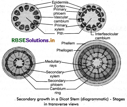
Explain the process of secondary growth in the stems of woody angiosperms with the help of schematic diagrams. What is its significance?
Answer:
The growth of the roots and stems in length with the help of apical meristem is called the primary growth. Apart from primary growth most dicotyledonous plants exhibit an .increase in girth. This increase is called the secondary growth. The tissues involved in secondary growth are the two lateral meristems: vascular cambium and cork cambium.
Vascular Cambium :
The meristematic layer that is responsible for cutting off vascular tissues - xylem and phloem is called vascular cambium. In the young stem it is present in patches as a single layer between the xylem and phloem, Later it forms a complete ring.
Formation of Cambial Ring :
In dicot stems, the cells of cambium present between primary xylem and primary phloem is the intrafascicular cambium. The cells of medullary rays, adjoining these intrafascicular cambium become meristematic and form the interfascicular cambium. Thus, a continuous ring of cambium is formed.
Activity of the Cambial Ring :
The cambial ring becomes active and begins to cut off new cells, both towards the inner and the outer sides. The cells cut off towards pith, mature into secondary xylem and the cells cut off towards periphery mature into secondary phloem. The cambium is generally more active on the inner side than on the outer. As a result, the amount of secondary xylem produced is more than secondary phloem and soon forms a compact mass. The primary and secondary phloems get gradually crushed due to the continued formation and accumulation of secondary xylem. The primary xylem however remains more or less intact, in or around the centre. At some places, the cambium forms a narrow band of parenchyma, which passes through the secondary xylem and the secondary phloem in the radial directions. These are the secondary medullary rays.

Question 4.
Draw illustrations to bring out anatomical difference between :
(a) Monocot root and Dicot root.
(b) Monocot stem and Dicot stem.
Answer:
(a)
|
Dicot Root |
Monocot Root |
|
1. Cortex is simple and homogenous. It is made up of parenchymatous cells. |
Cortex is well developed. In some cases it developes one or more layers of thick walled cells towards outer side called exodermis. Rest of the cells are parenchymatous. |
|
2. Endodermis is prominentwith casperian thickenings. |
Endodermis is more prominent showing casperian thickenings. |
|
3. Number or protoxylem group is less than six. |
Number of protoxylem groups are more than six (polyarch). |
|
4. Secondary growth takes place. |
There is no secondary growth. |
|
5. Metaxylem vessels are generally polygonal. |
Metaxylem vessels are generally oval or rounded. |
|
6. Conjunctive tissue is parenchymatous. |
Conjunctive tissue around metaxylem vessels is sclerenchymatous. |
|
7. Pith is very small or absent. |
Pith is large. |
(b)
|
Dicot Stem |
Monocot Stem |
|
1. Internally, the stem is differentiated into epidermis, cortex, endodermis, pericycle, stelar system and pith. |
The ground tissue is not differentiated into cortex, endodermis, pericycle and pith. |
|
2. Hypodermis is usually collenchymatous. |
Hypodermis is usually sclerenchymatous. |
|
3. Vascular bundles are arranged in a ring. |
Vascular bundles are scattered in the parenchymatous ground tissue. |
|
4. Parenchymatous medullary rays occur in between the vascular bundles. |
Medullary rays are absent. |
|
5. Bundle sheath is absent. |
Vascular bundles are surrounded by sclerenchymatous bundle sheath. |
|
6. Vascular bundles are conjoint, collateral or bicollateral, endarch and open. |
Vascular bundles are conjoint, collateral or concentric, endarch and closed. |
|
7. Vascular bundles are limited in number. |
Vascular bundles are numerous. |
|
8. Xylem vessels are generally polygonal in transverse section. |
Xylem vessels are generally oval in transverse section. |
|
9. Lysigenous cavity in xylem is absent. |
Lysigenous cavity is present in xylem. |
|
10. Phloem is composed of sieve tubes, companion cells and phloem parenchyma. |
Phloem is composed of sieve tubes and companion cells only. The phloem parenchyma are absent. |

Question 5.
Cut a transverse section of young stem of a plant from your school garden and observe it under the microscope. How would you ascertain whether it is a monocot stem or a dicot stem? Give reasons.
Answer:
During examination of a cross section of young stem if it shows following features it belongs to dicot stem :
- Internally, the stem is differentiated into epidermis, cortex vascular bundles and pith.
- Hypodermis, if present, it is collenchymatous.
- Vascular bundles are arranged in a ring.
- Vascular bundles are open.
During examination if the following features are seen, the young stem belongs to monocot.
- Internally, the stem is not differentiated into cortex and pith.
- Hypodermis, if present is sclerenchymatous.
- Vascular bundles are scattered in ground tissue.
- Each vascular bundle is closed and has a bundle sheath.
Question 6.
The transverse section of a plant material shows the following anatomical features, (a) the vascular bundles are conjoint, scattered and surrounded by a sclerenchymatous bundle sheaths (b) Phloem parenchyma is absent. What will you identify it as?
Answer:
It is a monocot stem.
Question 7.
Why are xylem and phloem called complex tissues?
Answer:
A group of more than one type of cells having common origin and working together as an unit is called complex tissue. Since both xylem and phloem are made up of several kinds of cells, they are called complex tissues.
Question 8.
What is stomatal apparatus? Explain the structure of stomata with a labelled diagram.
Answer:
The epidermal tissue system forms the outer-most covering of the whole plant body and comprises epidermal cells, stomata and the epidermal appendages - the trichomes and hairs. The epidermis is the outermost layer of the primary plant body. It is made up of elongated, compactly arranged cells, which form a continuous layer. Epidermis is usually single- layered. Epidermal cells are parenchymatous with a small amount of cytoplasm lining the cell wall and a large vacuole. The outside of the epidermis is often covered with a waxy thick layer called the cuticle which prevents the loss of water. Cuticle is absent in roots. Stomata are present in the epidermis of leaves. Stomata regulate the process of transpiration and gaseous exchange.
Each stomata is composed of two bean- shaped cells known as guard cells which enclose stomatal pore. In grasses, the guard cells are dumb-bell shaped. The outer walls of guard cells (away from the stomatal pore) are thin and the inner walls (towards the stomatal pore) are highly thickened. The guard cells possess chloroplasts and regulate the opening and closing of stomata. Sometimes, a few epidermal cells, in the’ vicinity of the guard cells become specialised in their shape and size and are known as subsidiary cells. The stomatal aperture, guard cells and the surrounding subsidiary cells are together called stomatal apparatus.

The cells of epidermis bear a number of hairs. The root hairs are unicellular elongations of the epidermal cells and help in absorb water and minerals from the soil. On the stem the epidermal hairs are called trichdmes. The trichomes in the shoot system are usually multicellular. They may be branched or unbranched and soft or stiff. They may even be secretory. The trichomes help in preventing water loss due to transpiration.

Question 9.
Name the three basic tissue systems in flowering plants. Give the tissue names under each system.
Answer:
Three basic tissues systems in flowering plants are:
1. Epidermal tissue system : It consists of
- Epidermis
- Cuticle and wax
- Stomata
- Trichomes.
2. Ground tissue system : It consists of
- Cortex
- Endodermis
- Pericycle
- Medullary rays
- Pith
- Ground tissue of leaves.
3. Vascular tissue system : It consists of
- Xylem
- Phloem.
Question 10.
How is the study of plant anatomy useful to us?
Answer:
The study of plant anatomy help in solving various problems related to taxonomy, phylogeny, food adulteration archeology and manufacture of various wood products.
Question 11.
What is periderm? How does periderm formation take place in the dicot stems?
Answer:
As the stem continues to increase in girth due to the activity of vascular cambium, the outer cortical and epidermis layers get broken and need to be replaced to provide new protective cell layers. To avoid such breaking of external tissues, the plant organs develop a secondary protective covering by replacing epidermis and other external tissues. The protective secondary tissues consisting of cork, cork cambium and secondary cortex, formed by the activity of secondary lateral meristem (the cork cambium), is called periderm.
The periderm consists of following three parts :
- Phellogen (the cork cambium)
- Phellem (cork)
- Phelloderm (the secondary cortex)
1. Phellogen (the cork cambium): The phellogen is a secondary meristem that arises from living permanent cells. The cells of phellogen appear almost rectangular and radially flattened in transverse section. They are thin walled, vacuolated, living cells with granular content and divide by periclinal divisions to give rise to phellem (cork) to outer side and phelloderm. (parenchymatous secondary cortex) towards inner side. The phellogen always remains single layered.
2. Phellem (Cork) : The cells cut off towards outer side from the phellogen may divide once or twice and then differentiate into phellem (cork). As they mature, they elongate tangentially and radially, develop secondary wall due to deposition of suberin over the primary wall of cellulose and becomes dead due to loss of living protoplasm. The suberin is a fatty substance which is deposited over the primary wall in the form of distinct lamellae. The secondary wall layer is impermeable to water and, therefore, protects the loss of water. The cells of cork are compactly arranged in radial rows, and act as water tight compartments.
3. Phelloderm (the secondary cortex) : The cells cut off towards inner side from the phellogen may also divide once or twice again and finally differentiate into phelloderm (secondary cortex). The cells of phelloderm are thin walled, contain living protoplasm and resemble the parenchymatous cells of primary cortex. The cells are loosely arranged with distinct intercellular spaces. In some plants, the cells develop ehloroplast and perform photosynthesis.

Question 12.
Describe the internal structure of a dorsiventral leaf with the help of labelled diagrams.
Answer:
The vertical section of a dorsiventral leaf through the lamina shows three main parts, namely, epidermis, mesophyll and vascular system. The epidermis which covers both the upper surface (adaxial epidermis) and lower surface (abaxial epidermis) of the leaf has a conspicuous cuticle. The abaxial epidermis generally bears more stomata than the adaxial epidermis. The latter may even lack stomata. The tissue between the upper and the lower epidermis is called the mesophyll. Mesophyll, which possesses chloroplasts and carry out photosynthesis, is made up of parenchyma. It has two types of cells - the palisade parenchyma and the spongy parenchyma.
The adaxially placed palisade parenchyma is made up of elongated cells, which are arranged vertically and parallel to each other. The oval or round and loosely arranged spongy parenchyma is situated below the palisade cells and extends to the lower epidermis. There are numerous large spaces and air cavities between these cells. Vascular system includes vascular bundles, which can be seen in the veins and the midrib. The size of the vascular bundles are dependent on the size of the veins. The veins vary in thickness in the reticulate venation of the dicot leaves. The vascular bundles are surrounded by a layer of thick walled bundle sheath cells.
