RBSE Solutions for Class 11 Biology Chapter 8 Cell: The Unit of Life
Rajasthan Board RBSE Solutions for Class 11 Biology Chapter 8 Cell: The Unit of Life Textbook Exercise Questions and Answers.
Rajasthan Board RBSE Solutions for Class 11 Biology in Hindi Medium & English Medium are part of RBSE Solutions for Class 11. Students can also read RBSE Class 11 Biology Important Questions for exam preparation. Students can also go through RBSE Class 11 Biology Notes to understand and remember the concepts easily.
RBSE Class 11 Biology Solutions Chapter 8 Cell: The Unit of Life
RBSE Class 11 Biology Cell: The Unit of Life Textbook Questions and Answers
Question 1.
Which of the following is not correct?
(a) Robert Brown discovered the cell.
(b) Schleiden and Schwann formulated the cell theory.
(c) Virchow explained that cells are formed from pre - existing cells.
(d) A unicellular organism carries out its life activities within a single cell.
Answer:
(a) Robert Brown did not discover the cell. The cell was discovered by Robert Hooke.

Question 2.
New cells generate from:
(a) Bacterial fermentation
(b) Regeneration of old cells
(c) Pre - existing cells.
(d) Abiotic materials.
Answer:
(c) According to biogenic theory, new cells always arise from pre - existing cells.
Question 3.
Match the following:
|
Column - I |
Column - II |
|
(a) Cristae |
(i) Flat membranous sacs in stroma |
|
(b) Cisternae |
(ii) Infoldings in mitochondria |
|
(c) Thylakoids |
(iii) Disc - shaped sacs in Golgi apparatus |
Answer:
|
Column - I |
Column - II |
|
(a) Cristae |
(ii) Infoldings in mitochondria |
|
(b) Cisternae |
(iii) Disc - shaped sacs in Golgi apparatus |
|
(c) Thylakoids |
(i) Flat membranous sacs in stroma |
Question 4.
Which of the following is correct:
(a) Cells of all living organisms have a nucleus.
(b) Both animal and plant cells have a well defined cell wall.
(c) In prokaryotes, there are no membrane bound organelles.
(d) Cells are formed de novo from abiotic materials.
Answer:
(c) In prokaryotes, there are no membrane bound organelles.
Question 5.
What is a mesosome in a prokaryotic cell? Mention the functions that it performs.
Answer:
Mesosome is a convoluted membranous structure formed in a prokaryotic cell by infolding the plasma membrane.
Functions:
- It bears respiratory enzymes and help in respiration.
- They help in synthesis of cell wall and replication of DNA.
Question 6.
How do neutral solutes move across the plasma membrane? Can the polar molecules also move across it in the same way? If not, then how are these transported across the membrane?
Answer:
- Neutral molecules move across the plasma membrane by simple passive diffusion.
- The movement of polar molecules across the non polar lipid bilayer requires carrier proteins. Carrier proteins are integral protein particles having certain affinity for specific solutes. As a result, they facilitate the transport of molecules across the membrane.
Question 7.
Name two cell - organelles that are double membrane bound. What are the characteristics of these two organelles? State their functions and draw labelled diagrams of both.
Answer:
Mitochondria and chloroplasts are the two organelles that are double membrane bound.
These are important cell organelles essential for aerobic respiration of eukaryotic cell. They are popularly called as “power house of cell” because they are the centre of release of energy. Mitochondria was first observed by Kolliker in 1880 when he was working on muscle cells of insects. Benda 1898 in named mitochondria.
Location : Mitochondria are found in nearly all eukaryotic cells. They vary in number and location according to cell type. A single mitochondrion is often found in unicellular organisms. Conversely, numerous mitochondria are found in human liver cells, with about 1000 to 2000 mitochondria per cell, making up 1/5 of the cell volume.
Structure : The size and shape of mitochondria varies. They are small rod shaped or spherical structures present in the cytoplasm of the cells which show aerobic respiration. The shape depends upon physiological condition of the cell.
Size: The size of the mitochondrion may vary from cell to cell. Usually, it is 0.2 p to 1.0 p in diameter, 2 p to 8 p in length.
Morphology : Each mitochondrion is bounded by two membranes, the outer and inner membranes which resemble the cell membrane and lipo-proteinic in nature. In between the two membranes, there is a space called perimitoehondrial space of 80 A width. The outer membrane- is 60-75 A thick that forms outer covering. The inner membrane is 50-70 A thick that forms numerous finger like or plate like infoldings into the cavity of mitochondrion called as cristae (sing = crista).
This cavity is filled with fine, granular mitochondrial matrix, which is gel-like and contains proteins, lipids, circular DNA molecules, 70s ribosomes. On the inner membrane and cristae under high resolving power of electron microscope studies have shown numerous stalked knob like projections, called as elementary particles or oxysomes or F0 -Fx particles or Racker’s particles. Each particle consists of a base, stalk and a head. They contain all enzymes involved in electron transport chain (ETC) and oxidative phosphorylation. Hence, these are also called as electron transport particles (ETP).
Chemical Composition : The chemical composition of mitochondria varies in different animal and plant cells. Enzymes of outer membranes: It contains enzymes that are involved in mitochondrial lipid synthesis, e.g., mono amine oxidase, kynurenine hydroxylase, fatty acids co-ligase etc.
Enzyme of inter membrane space : It contains several enzymes that use ATP passing out of the matrix to phosphorylate other nucleotides. The main enzymes are adenylate kinase and nucleoside diphosphokinase.
The term plastid was introduced by E. Haeckel in 1866. These are double membrane bounded, mostly pigment containing cytoplasmic organelles present in plant cells only. They are absent in fungal or animal cells. They are also present in some protists like Euglena. According to Schimper (1883), these are classified into three types : Leucoplasts, Chromoplasts, Chloroplasts.
1. Leucoplasts : These are colourless plastids found in the storage organs. They are found in large numbers in the cells of fruits, seeds, tubers etc. They are variously shaped i. e., oval, rod like or filamentous. They lack grana and photosynthetic pigments.
They are of three types : Amyloplasts, Aleuroplasts (Proteinoplast) and Elaioplasts or oleosomes.
- Amyloplasts: They store starch grains and found in potato tubers, rice, wheat etc.
- Aleuroplasts (Proteinoplasts): They store proteins and found in maize grains.
- Elaioplasts or Oleosomes: They store lipids and found in the endosperm of castor seeds.
2. Chromoplasts: These are formed either from chloroplasts or from leucoplasts. For example, green tomatoes and chillies turn red on ripening due to the formation of the red pigment called lycopin replacing the chlorophyll. Red pigment carotene is developed in the carrot by replacing leucoplast.
Chloroplast: Chloroplasts were first reported by Leeuwenhoek. They are found only in photosynthetic plants cells like mesophyll cells, guard cells and the cells of green algae. These are generally disc shaped with circular or oval elliptical outline. They may also be collar shaped (e.g., Ulothrix), ribbon shaped (e.g., Spirogyra), cup shaped (e.g., Chlamydomonas), stellate (e.g., Zyngnema), lens shaped, rounded or club shaped. Their number may vary from 1 per cell (e.g., Chlamydomonas, Chlorella, Ulothrix etc.) to 20-40 per cell (e.g., mesophyll cells).
Structure: The chloroplast is double membrane bound structure filled with a fluid matrix.
Question 8.
What are the characteristics of prokaryotic cells?
Answer:
Prokaryotic cells are those cell that does not possess membrane-bounded nucleus e.g., bacteria, cyanobacteria. The characteristics of prokaryotic cells are as follows:
- They are small in size 0.1 mm to 10 mm.
- They do not possess membrane-bound organelles.
- They have single circular DNA as genetic material and plasmid.
- They possess mesosomes for respiration.
- Some are autotrophic and some are saprotrophic.
Question 9.
Multicellular organisms have division of labour. Explain.
Answer:
Multicellular organisms are made up of several cells. All these cells perform specific functions. All the cells specialised for performing similar functions are grouped together as tissue in the body. Hence, a particular function is carried out by a group of cells at a definite place of the body. Similarly, different functions are carried out by different group of cells (tissues). This show division of labour in multicellular organisms.
Question 10.
Cell is the basic unit of life. Discuss in brief.
Answer:
Cell is called basic unit of life because its cytoplasm is the site of many biochemical reactions important for life. Hence, cells are called basic unit of life.
Question 11.
What are nuclear pores? State their function.
Answer:
Nuclear pores are tiny holes present in the nuclear membrane of the nucleus. They are formed by the fusion of two nuclear membranes. These holes allow specific substances to be transferred into a cell and out from it. both directions, between the nucleus and cytoplasm.
Question 12.
Both lysosomes and vacuoles are endomembrane structures, yet they differ in terms of their functions. Comment.
Answer:
Lysosomes are membrane bound vesicular structures holding a variety of enzymes such as lipases, proteases and carbohydrases. The purpose of lysosomes is to digest worn out cells. They are involved in the intracellular digestion of foreign food particles and microbes. Sometimes, they also acts as suicide bags.
On the other hand, vacuoles are storage sacs found in cells. They might store the waste products of cells. In unicellular organisms, the food vacuole contains the consumed food particles. It also plays a role in expelling excess water and some wastes from the cell.
Question 13.
Describe the structure of the following with the help of labelled diagrams:
(i) Nucleus
(ii) Centrosome.
Answer:
(i) Nucleus was discovered by an English biologist Robert Brown in 1831. It is important part of the cell, which controls all the cellular activities hence it is also called as “control centre” of the cell.
Occurence: The nucleus is present in all eukaryotic cells. Certain cells e.g., some sieve tube of vascular plants and the red blood cells of mammals lack nuclei at maturity. The cells deprived of nuclei cannot divide and differentiate. Prokaryotic cells do not have an organised nucleus with a nuclear envelope. Their genetic material is highly folded circular DNA molecule without nuclear membrane and called nucleoid.
On the basis of number of nuclei, a cell may be uninucleate (have a single nucleus), binucleate (having two nuclei) or mutlinucleate (having many nuclei). Some cells like the cells of skeletal muscles, becomes multinucleated by the fusion of cells. This multinucleated cells formed by such a manner is called syncytia (singular syncitium). The aseptate fungal hyphae in some fungi like Rhizopus lack the septa (or cross walls) and thus a continuous fluid filled multinucleate hyphae are formed. This condition is called coenocytic.
Shape and Size : The shape of nucleus may be rounded, cuboidal, oval, cylindrical, disc like, horse shoe shaped lobed. Lens shaped or even branched. Generally, the size of nucleus may vary from 5 to 25 pm in diameter depending upon cell type.
Structure: The structure includes following components:
- Nuclear envelope (karyotheca)
- Nucleoplasm
- Nucleolus
- Chromation
(ii) Occurence : Centrioles are found in almost all eukaryotic animal cells, protozoan protists (except some forms like Amoeba), some fungi and the cells of those eukaryotic plants where flagellate structures are present in the life cycle (like green algae, bryophytes, pteridophytes and cycads). They are absent in higher gymnosperms, some algae and fungi.
Central apparatus or Centrosome: Centrioles commonly occur in pairs. The pair of centrioles is often called diplosome. The distinctly staining region of cytoplasm surrounding the diplosome is called centrosphere or kinoplasm (= cytocentrum). This part is devoid of any other cell organelle. The complex formed of centrioles and centrosphere, is called centrosome or central apparatus.
Size : Centrioles are approximately 0.3-0.5 pm in length and 0.15 pm in diameter. They are visible under light microscope, but the details of centrioles structure were revealed only under electron microscope. Usually two centrioles are found associated together but at right angles to each other.
Ultra Structure : A centriole possesses a wheel of nine peripheral fibrils. Fibrils are absent in the centre. The arrangement is therefore called 9+0. Fibrils run parallel to one another but at an angle of 40°. Each fibril is made up of three sub fibres therefore, it is called triplet fibril. The three subfibres are in reality microtubules, joined together by their margins and therefore, sharing the common walls made of 2-3 protofilaments.
Question 14.
What is a centromere? How does the position of centromere form the basis of classification of chromosomes. Support your answer with a diagram showing the position of centromere on different types of chromosomes.
Answer:
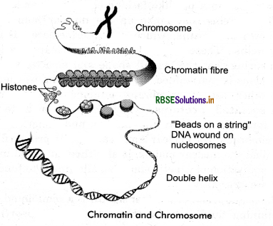
The chromosomes were first described by Strausberger in 1875. The term “chromosome” however was first used by Waldeyer in 1888. The chromatin fibres become condense to form short, thick, thread like structures called chromosomes during the dividing phase of cell cycle. Chemically the metaphase chromosomes contain 15-20% DNA, 10-15% RNA and 65-75% proteins.
A single human cell has approximately two meter long thread of DNA distributed among its forty six (23 pairs) chromosomes.
Structure of Chromosome : Typically a chromosome is made. up of two chromatids, a centromere and a secondary constriction. Sister chromatids are two identical copies of chromosome connected by a centromere. The two chromatids of one homologous chromosome with respect to those of the other homologue are called nonsister chromatids. The region where two sister chromatids of a chromosome appear to be joined during cell division is called centromere which is also termed as primary constriction. The chromosomes may have additional constrictions termed secondary constrictions near their ends. Parts of the chromosome beyond the secondary constriction is termed satellite. The protein structures on the sides of centromere where the spindle fibres attach during cell division to pull the chromosomes apart, are called kinetochore.
Types of Chromosomes : Each chromosome can be divided into two parts (p and q arms) with a constriction point called a centromere in the middle. However, the
centromere can be located in different positions and this forms the basis for the four different types of chromosome.
- Metacentric : When centromere is located in middle that means p and q arms are of comparable length {e.g., chromosomes 1, 3, 16, 19, 20).
- Submetacentric : When the centromere is located slightly away from the middle. In these chromosomes, p arm is shorter than q arm (e.g., chromosomes 2, 4,12,17, 18).
- Acrocentric : When the centromere is located close to the end of chromosome which form one extremely short (p arm) and one very long arm {q arm) e.g., chromosomes 13 to 15, 21, 22).
- Telocentric : When centromere is located at the end of chromosome that means one arm (p arm) is absent. This type of chromosomes are not found in humans.
A type of organelles that are found in both plant and animal cells. They were first discovered by Rhodin in 1954 in the mouse kindey cells.
Structure : A microbody is usually a vesicle with a spherical shape ranging from 0.2-0.5 micrometers in diameter. These occur in cytoplasm of a cell. They are surrounded by a single phospholipid bilayer membrane containing a matrix of intracellular material including enzymes and other proteins, but they do not seem to contain any genetic material to allow them to self replicate. The organelles in the microbody are peroxisome, glyoxysomes, glycosomes and hydrogenosomes. In vertebrates, microbodies are specially present in the liver and kidney.

- RBSE Solutions for Class 11 Biology Chapter 10 Cell Cycle and Cell Division
- RBSE Solutions for Class 11 Biology Chapter 9 Biomolecules
- RBSE Solutions for Class 11 Biology Chapter 7 Structural Organisation in Animals
- RBSE Solutions for Class 11 Biology Chapter 6 Anatomy of Flowering Plants
- RBSE Solutions for Class 11 Biology Chapter 5 Morphology of Flowering Plants
- RBSE Solutions for Class 11 Biology Chapter 4 Animal Kingdom
- RBSE Solutions for Class 11 Biology Chapter 3 Plant Kingdom
- RBSE Solutions for Class 11 Biology Chapter 2 Biological Classification
- RBSE Solutions for Class 11 Biology Chapter 1 The Living World
- RBSE Solutions for Class 11 Biology Chapter 5 पुष्पी पादपों की आकारिकी
- RBSE Class 11 Biology Important Questions Chapter 1 The Living World