RBSE Solutions for Class 11 Biology Chapter 21 Neural Control and Coordination
Rajasthan Board RBSE Solutions for Class 11 Biology Chapter 21 Neural Control and Coordination Textbook Exercise Questions and Answers.
Rajasthan Board RBSE Solutions for Class 11 Biology in Hindi Medium & English Medium are part of RBSE Solutions for Class 11. Students can also read RBSE Class 11 Biology Important Questions for exam preparation. Students can also go through RBSE Class 11 Biology Notes to understand and remember the concepts easily.
RBSE Class 11 Biology Solutions Chapter 21 Neural Control and Coordination
RBSE Class 11 Biology Neural Control and Coordination Textbook Questions and Answers
Question 1.
Briefly describe the structure of the following:
(a) Brain
(b) Eye
(c) Ear.
Answer:
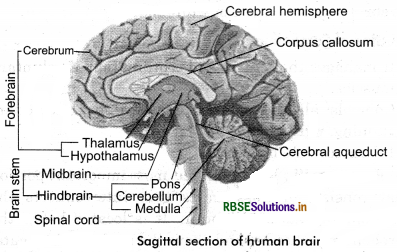
(a) Brain: The human brain is well protected by the skull. Inside the skull, the brain is covered by cranial meninges consisting of an outer layer called dura mater, a very thin middle layer called arachnoid and an inner layer (which is in contact with the brain tissue) called pia mater.
The brain can be divided into three major parts:
- Forebrain: The forebrain consists of the cerebrum, thalamus and hypothalamus. Functions including temperature, reproductive functions, eating, sleeping and the display of emotion.
- Midbrain: It is located between the thalamus/hypothalamus of the forebrain and pons of the hindbrain. The midbrain serves important functions in motor movement, particularly movements of the eye, and in auditory and visual processing.
- Hindbrain: It is hindbrain comprises pons, cerebellum and medulla. Controls functions outside conscious control, such as breathing and blood flow.
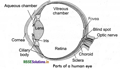
(b) Eye: The adult human eyeball is nearly a spherical structure. The wall of the eyeball is composed of three layers.
The external layer is composed of a dense connective tissue and is called the sclera.
The anterior portion of this layer is called the cornea. The middle layer, choroid, contains many blood vessels and looks bluish in colour.
The choroid layer is thin over the posterior two - thirds of the eyeball, but it becomes thick in the anterior part to form the ciliary body.
The ciliary body itself continues forward to form a pigmented and opaque structure called the iris which is the visible coloured portion of the eye.
The eyeball contains a transparent crystalline lens which is held in place by ligaments attached to the ciliary body. In front of the lens, the aperture surrounded by the iris is called the pupil whose diameter is regulated by the muscle fibres of iris. The inner layer is the retina and it contains three layers of neural cells from inside to outside - ganglion cells, bipolar cells and photoreceptor cells. There are two types of photoreceptor cells, namely, rods and cones. The daylight (photopic) vision and colour vision are functions of cones and the twilight (scotopic) vision is the function of the rods. The innermost ganglionic cells give rise to optic nerve fibre that forms optic nerve in each eye and is connected with the brain.
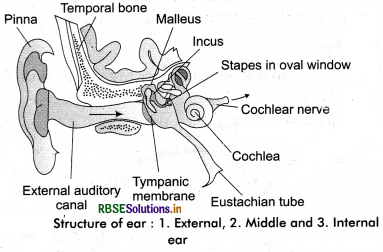
(c) Ear: It performs two sensory functions, hearing and maintenance of body balance. It can be divided into three major sections called the outer ear, the middle ear and the inner ear:
(i) Outer ear: It consists of the pinna and external auditory meatus (canal). The pinna collects the vibrations in the air which produce sound. The external auditory meatus leads inwards and extends up to the tympanic membrane (the eardrum). There are very fine hairs and wax - secreting glands in the skin of the pinna and the meatus. The tympanic membrane is composed of connective tissues covered with skin outside and with mucous membrane inside.
(ii) Middle ear: It contains three ossicles called malleus, incus and stapes which are attached to one another in a chain - like fashion. The malleus is attached to the tympanic membrane and the staps is attached to the oval window of the cochlea. The ear ossicles increase the efficiency of transmission of sound waves to the inner ear. A Eustachian tube connects the middle ear cavity with the pharynx. The Eustachian tube helps in equalizing the pressures on either side of the eardrum.
(iii) Inner ear: It is also known as the labyrinth. Labyrinth is divided into the bony labyrinth and a membranous labyrinth. Bony labyrinth is filled with perilymph while membranous labyrinth is filled with endolymph. The membranous labyrinth is divided into two parts - Vestibular apparatus and Cochlea.
The vestibular apparatus is composed of three semi-circular canals and the otolith (macula is the sensory part of saccule and utricle). The crista and macula are the specific receptors of the vestibular apparatus responsible for maintenance of the balance of the body and posture. The cochlea is a long and coiled outgrowth of sacculus. It is the main hearing organ. Cochlea consists of three membranes. The organ of Corti, a hearing organ, is located on the basilar membrane that has hair cells.

Question 2.
Compare the following:
(a) Central neural system (CNS) and peripheral neural system (PNS).
(b) Resting potential and action potential.
(c) Choroid and retina.
Answer:
(a) Central neural system (CNS) and peripheral neural system (PNS):
|
CNS |
PNS |
|
1. The coatral nervous system consists of (a) Brain and (b) Spinal cord. |
1. The peripheral nervous system consists of (a) cranial nervous and (b) spinal nervous, which connects the different parts of the body to CNS. |
|
2. It is protected by bony structure: Skull and spinal cord and meninges. |
2. It is surrounded by neurilemma. |
|
3. It has sensory and motor nerves along with connecting nerve cells. Which conduct the impulses between sensory and motor nerves. This controls and coordinates different responses. |
3. The sensory nerve, carry the stimulus to CNS and motor nerves carry the message to target organ: muscles or glands. |
(b) Resting potential and action potential:
|
Resting pontential |
Action potential |
|
1. The state of the nerve cell (neurolemma) with + charge on the outerside and - charge on the innerside is called resting potential. |
1. The state of the nerve cell (neurolemma) with - charge on the outerside and + charge on the inner side. It is called action potential, when a stimulus is given to it. |
|
2. The neurilemma is more permeable to Na+ than K+ ions. |
2. During this stage the neurilemma is highly permeable to Na+ and impermeable to K+ ions. |
|
3. Due to resting potential the sodium-potassium pump functioning the resting potential is formed. |
3. During this stage sodium- potassium pump does not function. Thus, the action potential is generated due to influx of Na+ ions in neuroplasm. |
|
4. During this stage the neuron does not conduct stimulus. |
4. During this stage the nerves conduct stimulus. |
(c) Choroid and Retina:
|
Choroid |
Retina |
|
1. It is the middle layer of the eye ball. |
1. It is the inner most layer of eye. |
|
2. It is made up of soft connective tissue. It has network of blood capillaries and branched pigmented. It is in contact with sclera and retina. |
2. It is a thin and soft layer. It is madeup of sensory and pigmented layers. Pigmented layer is in constant with choroid. |
|
3. Choroid layer separates from the sclera and forms iris. Due to the muscles present in it the size of pupil can be increased/ decreased. This works like a diaphragm of a camera. |
3. It has 2 types of photo sensitive cells: (a) Rod cells. (b) Cones cells. |
Question 3.
Explain the following processes:
(a) Polarisation of the membrane of a nerve fibre.
(b) Depolarisation of the membrane of a nerve fibre.
(c) Conduction of a nerve impulse along a nerve fibre.
(d) Transmission of a nerve impulse across a chemical synapse.
Answer:
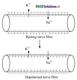
(a) Polarisation of the membrane of a nerve fibre:
During resting condition, the concentration of K+ ions is more inside the axoplasm while the concentration of Na+ ions is more outside the axoplasm. As a result, the potassium ions move faster from inside to outside as compared to sodium ions. Therefore, the membrane becomes positively charged outside and negatively charged inside. This is known as polarization of membrane or polarized nerve.
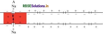
(b) Depolarisation of the membrane of a nerve fibre:
When an electrical stimulus is given to a nerve fibre, an action potential is generated. The membrane becomes permeable to sodium ions than to potassium ions. This results into positive charge inside and negative charge outside the nerve fibre. Hence, the membrane is said to be depolarized.
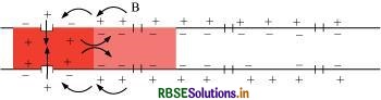
(c) Conduction of a nerve impulse along a nerve fibre:
There are two types of nerve fibres – myelinated and non-myelinated. In myelinated nerve fibre, the action potential is conducted from node to node in jumping manner. This is because the myelinated nerve fibre is coated with myelin sheath. The myelin sheath is impermeable to ions. As a result, the ionic exchange and depolarisation of nerve fibre is not possible along the whole length of nerve fibre. It takes place only at some point, known as nodes of Ranvier, whereas in non-myelinated nerve fibre, the ionic exchange and depolarization of nerve fibre takes place along the whole length of the nerve fibre. Because of this ionic exchange, the depolarized area becomes repolarised and the next polarized area becomes depolarized.
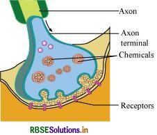
(d) Transmission of a nerve impulse across a chemical synapse:
Synapse is a small gap that occurs between the last portion of the axon of one neuron and the dendrite of next neuron. When an impulse reaches at the end plate of axon, vesicles consisting of chemical substance or neurotransmitter, such as acetylcholine, fuse with the plasma membrane. This chemical moves across the cleft and attaches to chemo-receptors present on the membrane of the dendrite of next neuron. This binding of chemical with chemo-receptors leads to the depolarization of membrane and generates a nerve impulse across nerve fibre. The chemical, acetylcholine, is inactivated by enzyme acetylcholinestrase. The enzyme is present in the post synaptic membrane of the dendrite. It hydrolyses acetylcholine and this allows the membrane to repolarise.
Question 4.
Draw the labelled diagrams of the following:
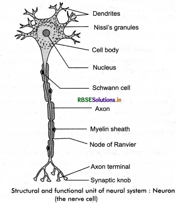
(a) Neuron
(b) Brain
(c) Eye
(d) Ear.
Answer:
(a) Neuron:
(b) Brain:
(c) Eye:
(d) Ear:
Question 5.
Write short notes on the following:
(a) Neural coordination
(b) Forebrain
(c) Mid - brain
(d) Hindbrain
(e) Retina
(f) Ear ossicles
(g) Cochlea
(h) Organ of Corti
(i) Synapse.
Answer:
(a) Neural coordination in human beings takes place due to the communication system of the body. It involves:
(i) Neural/nervous system and endocrinosystem. The nervous system consists of brain, spinal cord and nerves aring from these parts. These nerves collect information from out side the body and send to the CNS. Which sends the responses as messages to the muscles or glands and thus develops/establishes the neural coordination in the body for its response.
(b) Forebrain consists of the cerebrum, cerebral hemispheres, olfactory lobes and diencephalon (thalamus and hypothalamus). It helps in the interpretation of stimulus received by effector organs.
(c) Midbrain consists of tectum (visual and auditory stimuli) and tegmentum (contains nuclei for pain modulation, motor coordination and movement planning). It helps in the relay of impulse from effector organ to the forebrain.
(d) Pons, cerebellum and medulla together form the hindbrain. It helps to maintain the balance of body and body posture. It also has regulatory centres for controlling the involuntary actions.
(e) The retina is the innermost layer of the eyeball and contains photoreceptor rods and cones.
(f) Ear ossicles: Three small bones present between the tympanic membrane and oval window are collectively referred to as ossicles, they are namely malleus, incus and stapes. It increases the amplification of sound waves.
(g) Cochlea: Cochlea is a spiral hollow structure containing three fluid-filled canals. Organ of corti is located in middle cochlear canal and has hair cells (mechanoreceptors) on its basilar membrane. Thus, cochlea houses sensory system for hearing only and is not associated with balancing.
(h) Organ of corti: Organ of corti is located in middle cochlear canal and has hair cells (mechanoreceptors) on its basilar membrane. It generates the auditory impulse which is carried by auditory nerves.
(i) Synapse: Two neurons are never physically connected to each other and synapse is the region of close proximity between two neurons where information from one neuron is transmitted to the next one.
Question 6.
Give a brief account of:
(a) Mechanism of synaptic transmission.
(b) Mechanism of vision.
(c) Mechanism of hearing.
Answer:
(a) Synapse is a junction between two neurons. It is present between the axon terminal of one neuron and the dendrite of next neuron separated by a cleft.
There are two ways of synaptic transmission.
- Chemical transmission
- Electrical transmission
1. Chemical transmission: When a nerve impulse reaches the end plate of the axon, it releases a neurotransmitter (acetylcholine) across the synaptic cleft. This chemical is synthesized in the cell body of the neuron and is transported to the axon terminal. The acetylcholine diffuses across the cleft and binds to the receptors present on the membrane of next neuron. This causes depolarization of membrane and initiates an action potential.
2. Electrical transmission: In this type of transmission, an electric current is formed in the neuron. This electric current generates an action potential and leads to transmission of a nerve impulse across the nerve fibre. This represents a faster method of nerve conduction than the chemical method of transmission.
(b) Mechanism of vision:
Retina is the innermost layer of the eye. It contains three layers of cells – inner ganglion cells, middle bipolar cells, and outermost photoreceptor cells. A photoreceptor cell is composed of a protein called as opsin and an aldehyde of vitamin A called as retinal. When light rays are focused on the retina through the cornea, it leads to the dissociation of retinal from opsin protein. This changes the structure of opsin. As the structure of opsin changes, the permeability of membrane changes, generating a potential difference in the cells. This generates an action potential in the ganglionic cells and is transmitted to the visual cortex of the brain via optic nerves. In the cortex region of the brain, the impulses are analysed and the image is formed on the retina.
(c) Mechanism of hearing:
The pinna of the external region collects the sound waves and directs it towards ear drum or external auditory canal. These waves strike the tympanic membrane and vibrations are created. Then, these vibrations are transmitted to the oval window, fenestra ovalis, through three ear ossicles, named as malleus, incus, and stapes. These ear ossicles act as a lever and transmit the sound waves to internal ear. These vibrations from fenestra ovalis are transmitted into the cochlear fluid. This generates sound waves in the lymph. The formation of waves generates a ripple in the basilar membrane. This movement bends the sensory hair cells present on the organ of Corti against tectorial membrane. As a result of this, sound waves are converted into nerve impulses. These impulses are then carried to the auditory cortex of brain via auditory nerves. In cerebral cortex of the brain, the impulses are analysed and the sound is recognized.
Question 7.
Answer briefly:
(a) How do you perceive the colour of an object?
(b) Which part of our body helps us in maintaining the body balance?
(c) How does the eye regulate the amount of light that falls on the retina?
Answer:
(a) The colour of the object is analysed by the three types cones cells present in the retina. The light when falls on these cone cells they get stimulated and hence, we can see the colour.
(b) The internal ear cochlea helps in maintaining the body balance.
(c) The contraction in unstriped muscles and circular sphincter muscles of iris regulate the pupil - aperture that helps to regulate the amount of light reflex on the retina.
Question 8.
Explain the following:
(a) Role of Na+ in the generation of action potential.
(b) Mechanism of generation of light induced impulse in the retina.
(c) Mechanism through which a sound produces a nerve impulse in the inner ear.
Answer:
(a) Sodium ions play an important role in the generation of action potential. When a nerve fibre is stimulated, the membrane potential decreases. The membrane becomes more permeable to Na+ ions than to K+ ions. As a result, Na+ diffuses from the outside to the inside of the membrane. This causes the inside of the membrane to become positively - charged, while the outer membrane gains a negatively charge. This reversal of polarity across the membrane is known as depolarisation. The rapid inflow of Na+ ions causes the membrane potential to increase, thereby generating an action potential.
(b) Retina is the innermost layer of the eye. It contains three layers of cells – inner ganglion cells, middle bipolar cells, and outermost photoreceptor cells. Photoreceptor cells are composed of a protein called opsin and an aldehyde of vitamin A called retinal. When light rays are focused on the retina through the cornea, retinal gets dissociated from opsin. As a result, the structure of opsin gets changed. This in turn causes the permeability of the membrane to change, thereby generating a potential difference in the cells. Consequently, an action potential is generated in the ganglion cells and is transmitted to the visual cortex of the brain via the optic nerves. In the cortex region of the brain, the impulses are analysed and the image is formed on the retina.
(c) The pinna of the external ear collects the sound waves and directs them to the tympanic membrane (ear drum) via the external auditory canal. The ear drum then vibrates the sound waves and conducts them to the internal ear through the ear ossicles. The ear ossicles increase the intensity of the sound waves. These vibrating sound waves are conducted through the oval window to the fluid in the cochlea. Consequently, a movement is created in the lymph. This movement produces vibrations in the basilar membrane, which in turn stimulate the auditory hair cells. These cells generate a nerve impulse, conducting it to the auditory cortex of the brain via afferent fibres. The auditory cortex region interprets the nerve impulse and sound is recognised.
Question 9.
Differentiate between:
(a) Myelinated and non - myelinated axons.
(b) Dendrites and axons.
(c) Rods and cones.
(d) Thalamus and Hypothalamus.
(e) Cerebrum and cerebellum.
Answer:
(a) Difference between
|
Myelinated axon |
Non - myelinated axons |
|
1. The axon (fibre) having a myelin sheath on the neurolemma are called myelinated axon. |
1. The axon (fibre) lacking a myelin sheath is called non - myelinated axon. |
|
2. These axon fibres have schwann cells attached to it that produce myelin. It is made up of fats and appear white. |
2. These axon fibres do not have schwann cells. The axons are free of them and appear grey. |
|
3. They provide an insulation to the axon and the transmission/conduction of impulse. |
3. Due to no insulation the conduction of impulse is slow. |
|
4. Such axons are found in CNS. |
4. Such axons are present in ANS. |
|
5. They form the white matter of the CNS. |
5. They form the grey matter of the CNS. |
(b) Dendrites and Axon
|
Dendrites |
Axon |
|
1. They emerge from the cell body or cyton of the neuron. These are - usually small, thick at baselorigin and thin and branched at tip. |
1. They emerge from the cell body and are thick, long, myelinated or non myelinated and branched at the terminal end. |
|
2. Their number is - one (bipolar) and many (multi polar). |
2.It is one in number. |
|
3. They conduct the stimulus received towards the cyton. |
3.It conducts the impulse generated as a result of action potential due to changes in Na+ and K+ ions on the neurolemma. |
(c) Rods and Cones
|
Rods |
Cones |
|
1. These are rod shaped cells of retina. |
1. These are cone shaped cells of retina. |
|
2. These cells differentiate between light and dark thus are twilight (scotopic) photo sensitive. |
2. These cells are of three types. They are sensitive to day light (photopic) and distinguish colours. (for colour vision). |
|
3. They have rhodopsin or visual purple a purplish protein or derivative of vitamin A. |
3. They have lodopsin. |
|
4. They become active in less light or dark. |
4. They are active/functional only in day light. |
|
5. Due to less number they causes night blindness. |
5. Due to the less number they causes colour blindness. |
(d) Thalamus and Hypothalamus
|
Thalamus |
Hypothalamus |
|
1. It is surroundedlcovered by cerebrum. |
1. It is located at the base of thalamus. |
|
2. It is madeup of grey matter and appear like lobes. It consists of upper portion of the posterior portion diencephalon. |
2. It is the basal portion of the posterior portion of diencephalon. |
|
3. The group of neurons form thermo regulation centre here. |
3. The neurons are with large nuclei. The neurons form four main portions. |
|
4. It is the relay centre for heat, pain, touch, shivering, hearing, vision or sensory information. |
4. It regulates the experiences shunger, thirst, satisfaction, anger, excitment, sexual pleasure etc. |
(e) Cerebrum and cerebellum
|
Cerebrum |
Cerebellum |
|
1. It is the main part of the fore brain. |
1. It is the main part of the hind brain. |
|
2. It is having two cerebral hemispheres that are attachediconnected by corpus callosum. |
2. It has two occipital hemispheres on both right and left sides. Which are connected by vermis. |
|
3. The cavity of the cerebral hemispheres are called posterior ventricles. |
3. It does not have cavities. |
|
4. It is the centre of 98% mental function - such as intelligencelknowledge, desire, voluntary actions, rememberance, voice,thinking etc. |
4. It helps to keep the body posture and balancing. It coordinates the muscular activities. |
|
5. It regulates both voluntary and involuntary actions. |
5. Its regulation is involuntary. |
Question 10.
Answer the following:
(a) Which part of the ear determines the pitch of the sound?
(b) Which part of the human brain is the most developed?
(c) Which part of our central neural system acts as a master clock.
Answer:
(a)The cochlea has organ or corti is responsible for determining the pitch of the sound.
(b) Cerebrum is the most developed part of the human brain.
(c) Brain acts as the master clock in our body.
Question 11.
The region of the vertebrate eye, where the optic nerve passes out of the retina, is called the:
(a) Fovea
(b) Iris
(c) Blind spot
(d) Optic Chiasma.
Answer:
(c) Blind spot.
Question 12.
Distinguish between:
(a) Afferent neurons and efferent neurons.
(b) Impulse conduction in a myelinated nerve fibre and unmyelinated nerve fibre.
(c) Aqueous humor and vitreous humor.
(d) Blind spot and yellow spot.
(e) Cranial nerves and spinal nerves.
Answer:
(a) Afferent nerves and efferent nerves
|
Afferent Nerves |
Efferent Nerves |
|
1. Afferent nerves arise from periphery and transmit the stimuli to CNS. |
1. Efferent nerves arise from the CNS and carry the message to the effective organs. |
|
2. These are also called sensory nerves. |
2. These are also called motor nerves. |
|
3. These are unipolar nerves. |
3. These are multicellular nerves. |
(b) Impulse conduction in a myelinated fibre and unmyelinated nerve fibre
|
Impulse conduction in a Myelinated fibre |
Impulse conduction in a Unmyelinated/non myelinated fibre |
|
1. The impulse conduction is almost 10 times faster than the non myelinated fibre. |
1. The impulse conduction is slower than myelinated fibre. |
|
2. The impulse jumps from one node of ranvier node to another. Hence, the speed is faster. |
2. The impulse travels along the length of axon hence, it is slower. |
|
3. It utilizes less energy. |
3. It needs, comparatively more energy. |
(c) Aqueous humor and Vitreous humor
|
Aqueous Humor |
Vitreous Humor |
|
1. The liquid/fluid material present in between the cornea and lens is called aqueous humor. |
1. It is the material present in between the lens and retina called vitreous humor. |
|
2. It is liquid in nature and nutrients and O2 present.It removes the exeretory materials. |
2. It is thick/gel/jelly like substance and has - water, salts, etc., and their collagen fibres. |
|
3. It maintains the pressure on lens. |
3. It maintains the shape and pressure of the eye ball. |
(d) Bind spot and Yellow spot
|
Blind Spot |
Yellow Spot |
|
1. The spot of retina that lacks rod and cones cells is called bind spot. |
1. The spot or retina that has concentration of cone cells is called yellow spot. |
|
2. It does not have any pigments and hence, it does not form any image. |
2. It has yellow pigment the image in most accurate. |
|
3. The optic nerve emerges out from here and no image is formed. |
3. It is present on the mid of the retina at the posterior portion. |
|
4. It is insensitive to light. |
4. It is photosensitive. |
|
5. It has nerve cells and blood vessels near it. |
5. It lacks nerve cells and blood vessels. |
(e) Cranial nerves and Spinal Nerves
|
Cranial Nerves |
Spinal Nerves |
|
1. These emerge from the brain. |
1. These emerge from the spinal cord. |
|
2. There are 12 pairs of cranial nerves. |
2. There are 31 pairs of spinal nerves. |
|
3. They are categorised as: (a) Sensory: I, II, and VIII (b) Motor: III, IV and IV (c) Mixed: VII, IX, X, XI and XII. |
3. They are: Posterior sensory nerves and anterior motor nerves. Each spinal nerve divides into: posterior, anterior and mixed type. |

- RBSE Solutions for Class 11 Biology Chapter 10 Cell Cycle and Cell Division
- RBSE Solutions for Class 11 Biology Chapter 9 Biomolecules
- RBSE Solutions for Class 11 Biology Chapter 8 Cell: The Unit of Life
- RBSE Solutions for Class 11 Biology Chapter 7 Structural Organisation in Animals
- RBSE Solutions for Class 11 Biology Chapter 6 Anatomy of Flowering Plants
- RBSE Solutions for Class 11 Biology Chapter 5 Morphology of Flowering Plants
- RBSE Solutions for Class 11 Biology Chapter 4 Animal Kingdom
- RBSE Solutions for Class 11 Biology Chapter 3 Plant Kingdom
- RBSE Solutions for Class 11 Biology Chapter 2 Biological Classification
- RBSE Solutions for Class 11 Biology Chapter 1 The Living World
- RBSE Solutions for Class 11 Biology Chapter 5 पुष्पी पादपों की आकारिकी