RBSE Solutions for Class 11 Biology Chapter 18 Body Fluids and Circulation
Rajasthan Board RBSE Solutions for Class 11 Biology Chapter 18 Body Fluids and Circulation Textbook Exercise Questions and Answers.
Rajasthan Board RBSE Solutions for Class 11 Biology in Hindi Medium & English Medium are part of RBSE Solutions for Class 11. Students can also read RBSE Class 11 Biology Important Questions for exam preparation. Students can also go through RBSE Class 11 Biology Notes to understand and remember the concepts easily.
RBSE Class 11 Biology Solutions Chapter 18 Body Fluids and Circulation
RBSE Class 11 Biology Body Fluids and Circulation Textbook Questions and Answers
Question 1.
Name the components of the formed elements in the blood and mention one major function of each of them.
Answer:
The blood is a fluid connective tissue made of plasma (55%) and blood corpuscles (45%). The formed elements are cells and cell fragments suspended in the plasma. The three classes of formed elements are the red blood corpuscles (RBCs) or erythrocytes, white blood corpuscles (WBCs) or leucocytes and blood platelets.
Functions:
- RBCs: They play a significant role in the transportation of respiratory gases.
- WBCs: They help to fight against the infections and are responsible for the immune system in the body.
- Platelets: They help in coagulation of blood.

Question 2.
What is the importance of plasma proteins?
Answer:
Importance of Plasma Proteins: The blood contains about 7-8% plasma, proteins. Main plasma proteins are albumins, globulins, prothrombin and fibrinogens. They are formed in liver. Blood proteins are globular and soluble. These found in the form of calloids.
- Albumin proteins maintain osmotic pressure of the blood.
- Globulin proteins are of three types: alpha (α), beta (ß) and gamma (δ). Alpha and beta globulins transport proteins to different parts of the body, while gamma globulins protects the body from infections.
- Prothrombin and fibrinogen help in coagulation of blood.
Question 3.
Match column I with column II.
|
Column - I |
Column - II |
|
(a) Eosinophils |
(i) Coagulation |
|
(b) RBC |
(ii) Universal recipient |
|
(c) AB Group |
(iii) Resist infections |
|
(d) Platelets |
(iv) Contraction of heart |
|
(e) Systole |
(v) Gas transport |
Answer:
|
Column - I |
Column - II |
|
(a) Eosinophils |
(iii) Resist infections |
|
(b) RBC |
(v) Gas transport |
|
(c) AB Group |
(ii) Universal recipient |
|
(d) Platelets |
(i) Coagulation |
|
(e) Systole |
(iv) Contraction of heart |
Question 4.
Why do we consider blood as a connective tissue?
Answer:
Like the other connective tissues, blood is also mesodermally derived and has an extra - cellular matrix called plasma. It connects different body systems and takes part in the transportation of oxygen and various nutrients inside the body and removal of the waste materials out of the body. Hence, blood is considered as a connective tissue.
Question 5.
What is the difference between lymph and blood?
Answer:
Differences between Lymph and Blood:
|
Lymph |
Blood |
|
1. It consists of plasma and leucocytes (lymphocytes most abundant). |
1. It consists of plasma, erythrocytes, leucocytes and platelets. |
|
2. It is colourless as haemoglobin is absent. |
2. It is red in colour due to presence of haemoglobin in erythrocytes. |
|
3. Its plasma has fewer proteins and less calcium and phosphorus. |
3. Its plasma has more proteins, calcium and phosphorus. |
|
4. It transfers materials from the blood to the body cells and vice versa, therefore it acts as a ‘middle man’. |
4. It carries materials towards and away from the tissue, therefore, it acts as a ‘vehicle’. |
Question 6.
What is meant by double circulation? What is its significance?
Answer:
Double circulation: In human and other mammals there are two circuits of blood circulation for greater efficiency and to completely prevent the mixing of oxygenated and deoxygenated blood. Usually it is called double circulation.
- First circuit: Systemic circulation: It involves the circulation of oxygenated blood from the left ventricle of the heart to the aorta. Then a network of arteries, aerials and capillaries supplies this oxygenated blood to various tissues in the body from tissues, the deoxygenated blood is collected by system of venules, veins and vena cava and emptied into the right atrium.
- Second circuit: Pulmonary circulation: It involves the circulation of deoxygenated blood from right ventricle to the pulmonary artery, which then carries blood to the lungs for oxygenation. Then the oxygenated blood from lungs, is carried by the pulmonary veins into the left atrium.
Significance of double circulation: In double circulation there is complete separation of oxygenated and deoxygenated blood which allows a more efficient supply of oxygen to the body cells.
Question 7.
Write the differences between:
(a) Blood and lymph
(b) Open and closed system of circulation
(c) Systole and Diastole
(d) P - wave and T - wave.
Answer:
(a) Differences between blood and lymph:
|
Lymph |
Blood |
|
1. It consists of plasma and leucocytes (lymphocytes most abundant). |
1. It consists of plasma, erythrocytes, leucocytes and platelets. |
|
2. It is colourless as haemoglobin is absent. |
2. It is red in colour due to presence of haemoglobin in erythrocytes. |
|
3. Its plasma has fewer proteins and less calcium and phosphorus. |
3. Its plasma has more proteins, calcium and phosphorus. |
|
4. It transfers materials from the blood to the body cells and vice versa, therefore it acts as a ‘middle man’. |
4. It carries materials towards and away from the tissue, therefore, it acts as a ‘vehicle’. |
(b) Differences between open and closed system of circulation:
|
Open System of Circulation |
Closed System of Circulation |
|
1. In this system, blood pumped by the heart passes through large vessels into open spaces or body cavities called sinuses. |
1. In this system, blood pumped by the heart passes through closed network of blood vessels. |
|
2. Blood is in direct contact with the tissue cells. |
2. Blood does not come in direct contact with the tissue cells. |
|
3. Exchange of materials occurs directly between blood and tissue cells. |
3. Exchange of materials between tissue cells and blood occurs via tissue fluid. |
|
4. Blood flow very slow. |
4. Blood flow is quite rapid. |
|
5. Respiratory pigment if present, is dissolved in the plasma, no RBCs are present. |
5. Respiratory pigment is present and may be dissolved in the plasma but is usually held in RBCs. |
|
6. Occurs in arthropods and most molluscs. |
6. Occurs in annelids and vertebrates. |
(c) Differences Between Systole and Diastole:
|
Systole |
Diastole |
|
1. The contraction of the heart muscles is called systole. |
1. The relaxation of the heart muscles is called Diastole. |
|
2. It causes a decrease in the volume of the heart chambers and forces the blood out of them. |
2. It brings the heart chambers back to their original size to receive more blood. |
(d) Differences Between P - wave and T - wave:
|
P - Wave |
T - Wave |
|
1. P - wave marks the electrical excitation of atria, i.e., depolarization of atria. |
1. T - wave marks the return of ventricles from excited state to normal state, i.e., ventricular repolarisation. |
|
2. It causes the contraction of both the atria. |
2. It causes the relaxation of the ventricle. |
Question 8.
Describe the evolutionary change in the pattern of heart among the vertebrates.
Answer:
A comparative study of the structure of hearts of vertebrates reveals that their is an evolutionary change in the pattern of heart among the vertebrates. All vertebrates possess a muscular chambered heart. The heart of vertebrates is originated from embryonic mesoderm. In embryonic stage, two longitudinal endothelial canals in basal mesentry beneath archenteron, unite to form heart. The heart collects the blood from body and pumps it to body parts through arteries. In vertebrates there four types of hearts are found.
1. Single Chambered Heart: The members of cephalochordate have single - chambered heart. In these animals, the aorta present beneath pharynx becoming muscular to pumps blood, it is called single - chambered heart.
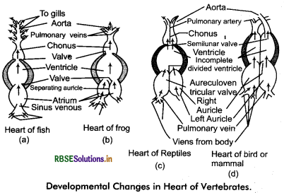
2. Two Chambered Heart: This type of heart is found in fishes. This heart pumps the deoxygenated blood into gills. Being oxygenated blood from gills then distributed into body parts. It has one auricle and one ventricle. This type of heart is called venous heart.
3. Three Chambered Heart: This type of heart is found in amphibians. This type of heart has three chambers two auricles and a ventricle. Sinus venosus opens on the dorsal plane of right auricle. Left auricle contains pure blood and right auricle contains impure blood. In amphibians the mixed blood is pumped by aorta to different organs of the body. Pulmonary artery carries the blood into lungs. In this blood circulation is unidirectional.
4. Four Chambered Heart: Mostly reptiles have two auricles and two incompletely divided ventricles. The heart of crocodile has two auricles and two ventricles. The heart of birds and mammals have four chambers two auricles and two ventricles. The left auricle and left ventricle contain oxygenated blood it is pumped into body parts through systemic arch. Right auricle collects deoxygenated blood which is pumped by right ventricle to lungs for oxygenation. Therefore, the left part of the heart is called pulmonary heart and the right part of the heart is called systemic heart. In these animals double circulation is found. There is no possibility to mixing blood in this type of circulation.
Question 9.
Why do we call our heart myogenic ?
Answer:
The Heart of Human is Myogenic:
The wall of the heart is made of cardiac muscles. These muscles are like striped muscles in structure but their function is involuntary like unstriped muscles. Heart beat originates through special muscular tissue in this type of heart. This tissue does not have contractibility but it has the ability to generate motor impulse by self excitation and transmit it as cardiac impulse. Molluscs and vertebrates have myogenic heart. In this type of heart, neural and hormonal stimuli do not generate cardiac impulse (heart beat) but affect the rate of heart beat. Therefore, heart beat continues even if the nervous connection of heart is removed. Due to this quality the heart of human is called myogenic.
Question 10.
Sino - atrial node is called the pacemaker of our heart. Why?
Answer:
The Sino - atrial (SA) node is a specialized bundle of neurons that have the ability to generate action potential without any external stimuli. This action potential is responsible for initiating and maintaining the rhythmic contraction of the heart. Due to this ability the SA. node is called the pacemaker.
Question 11.
What is the significance of atrio - ventricular node and atrio - ventricular bundle in the functioning of heart?
Answer:
Atrio - ventricular node (AVN) is a mass of neuromuscular tissue, which is situated in wall of right atrium, near the base of inter - atrial septum. AV node is the pace setter of the heart, as it transmits the impulses initiated by SA node to all parts of ventricles. Atrio-ventricular bundle (AV bundle) or bundle of His is a mass of specialized fibers which originate from the AVN. Within the myocardium of the ventricles the branches of bundle of His divided into a network of fibres called Purkinje fibres convey impulse of contraction from the AVN to the myocardium of the ventricle.
Question 12.
Define a cardiac cycle and the cardiac output.
Answer:
Cardiac Cycle: The sequential events in the heart which are repeated cyclically is called cardiac cycle and it consists of systole (contraction) and diastole (relaxation) of both the atria and ventricles. The duration of a cardiac cycle is 0.8 seconds. Periods of cardiac cycle are atrial systole (0.1 second), ventricle systole (0.3 second) and complete cardiac diastole (0.4 second).
Cardiac output: The amount of blood pumped by heart per minute is called cardiac output. It is calculated by multiplying stroke volume (volume of blood pumped by each ventricle per minute) with heart rate (number of beats per minute). The heart of normal person beats 72 times per minute and pumped out about 70 mL of blood per beat. Therefore, cardiac output averages 5000 mL or 5 L.
Question 13.
Explain heart sounds.
Answer:
Heart sounds: The beating of heart produces characteristic sounds which can be heard by using stethoscope. In a normal person, two sounds are produced per heart beat. The first heart sound ‘Lubb’ is low pitched, not very loud and of long duration. It is caused partly by the closure of the bicuspid and tricuspid valves and partly by the contraction of muscles in the ventricles. The second heart sound ‘Dub’ is high pitched, louder, sharper and shorter in duration. It is caused by the closure of the semilunar valves and marks the end of ventricular systole.
Question 14.
Draw a standard ECG and explain the. different segments in it.
Answer:
Electrocardiography (ECG): This is the graphical representation of the cardiac cycle as produced by the electrograph. The diagrammatic representation of a standard ECG is shown below.
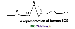
Each peak in ECG represented by the letters P to T, corresponds to a specific electrical activity of the heart.
- First peak ‘P’ indicates atrial depolarization (atrial contraction).
- Next QRS complex represents the depolarization of the ventricles (ventricular contraction).
- The last T - wave represents the ventricular repolarization (ventricular relaxation).

- RBSE Solutions for Class 11 Biology Chapter 10 Cell Cycle and Cell Division
- RBSE Solutions for Class 11 Biology Chapter 9 Biomolecules
- RBSE Solutions for Class 11 Biology Chapter 8 Cell: The Unit of Life
- RBSE Solutions for Class 11 Biology Chapter 7 Structural Organisation in Animals
- RBSE Solutions for Class 11 Biology Chapter 6 Anatomy of Flowering Plants
- RBSE Solutions for Class 11 Biology Chapter 5 Morphology of Flowering Plants
- RBSE Solutions for Class 11 Biology Chapter 4 Animal Kingdom
- RBSE Solutions for Class 11 Biology Chapter 3 Plant Kingdom
- RBSE Solutions for Class 11 Biology Chapter 2 Biological Classification
- RBSE Solutions for Class 11 Biology Chapter 1 The Living World
- RBSE Solutions for Class 11 Biology Chapter 5 पुष्पी पादपों की आकारिकी