RBSE Class 11 Biology Important Questions Chapter 20 Locomotion and Movement
Rajasthan Board RBSE Class 11 Biology Important Questions Chapter 20 Locomotion and Movement Important Questions and Answers.
Rajasthan Board RBSE Solutions for Class 11 Biology in Hindi Medium & English Medium are part of RBSE Solutions for Class 11. Students can also read RBSE Class 11 Biology Important Questions for exam preparation. Students can also go through RBSE Class 11 Biology Notes to understand and remember the concepts easily.
RBSE Class 11 Biology Chapter 20 Important Questions Locomotion and Movement
Multiple Choice Questions
Question 1.
Hydra captures its prey by:
(a) cilium
(b) flagellum
(c) tentacles
(d) setae
Answer:
(c) tentacles

Question 2.
Which of these are present in cell:
(a) microtubules
(b) microfflaments
(c) Both (a) and (b)
(d) Only (a)
Answer:
(c) Both (a) and (b)
Question 3.
Tick (√) the movement absent in human being cells is:
(a) amoeboid
(b) ciliary
(c) muscular
(d) tentacular
Answer:
(d) tentacular
Question 4.
Amoeboid movement in cells is by formation of:
(a) psuedo
(b) podium
(c) psuedopodium
(d) Amoeba
Answer:
(c) psuedopodium
Question 5.
The clearing/removal of dust particles and foreign particles inhaled in trachea is removed by:
(a) amoeboid movement
(b) ciliary movement
(c) tentacular movement
(d) sneezing
Answer:
(b) ciliary movement
Question 6.
The locomotory organ tube feet is of:
(a) fishes
(b) annelids
(c) starfish
(d) earthworms
Answer:
(b) annelids
Question 7.
What is the origin of muscles form in the embryo?
(a) Ectoderm
(b) Endoderm
(c) Mesoderm
(d) Embryo
Answer:
(c) Mesoderm
Question 8.
On the basis of location of muscles in our body it can be categorised into:
(a) one type
(b) three types
(c) two types
(d) four types
Answer:
(c) two types
Question 9.
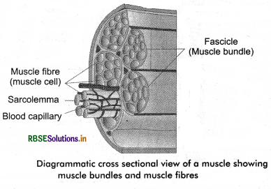
The labelled structures are:
(a) fasciculi and muscle fibre
(b) muscle fibre and fasciculi
(c) cell and nucleus
(d) bundle and Filament
Answer:
(a) fasciculi and muscle fibres
Question 10.
Which muscles present in your body can be controlled by your will?
(a) Cardiac
(b) Visceral
(c) Skeletal
(d) Smooth
Answer:
(c) Skeletal
Question 11.
In which manner the muscle fibres are arranged in skeletal muscles?
(a) Bundles
(b) Isolated
(c) Separated
(d) None of these
Answer:
(a) Bundles
Question 12.
The property of a cell to have more than one nucleus per cell is called:
(a) unicellular
(b) bicellular
(c) syncytium
(d) None of these
Answer:
(c) syncytium
Question 13.
Identify the type of ‘ions’ found in sarcoplasmic reticulum:
(a) K+
(b) Na+
(c) Ca++
(d) Cl+
Answer:
(c) Ca++
Question 14.
The ‘I’ - band of skeletal muscle myofilament is mark of:
(a) protein
(b) actin
(c) myosin
(d) troponin
Answer:
(b) actin
Question 15.
The ‘thick rod’ like myofilament in myofilament is of:
(a) protein
(b) actin
(c) myosin
(d) troponin
Answer:
(c) myosin
Very Short Answer Type Questions
Question 1.
Name an unicellular organism and location in human body that has cilia.
Answer:
(a) Paramoecium
(b) Oviduct.
Question 2.
Which locomotory organs are present in human?
Answer:
Limbs: Hind limbs (legs).
Question 3.
What are the purposes of locomotion in organisms?
Answer:
The purpose of locomotion in organisms are: search of food, shelter, mate and escaping from extreme climatic conditions.
Question 4.
Complete the box:
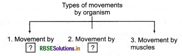
Answer:
- Cellular organelles.
- Body appendages.
Question 5.
Give an example of flagellar movement in human body.
Answer:
Movement of spermatozoan in the female reproductive tract.
Question 6.
Mention the properties in muscles.
Answer:
- Excitability
- Elasticity
- Extensibility
- Contractibility.
Question 7.
Mention the other names of skeletal muscles.
Answer:
Other names are: Fast muscles, voluntary muscles, striped muscles and striated muscles.
Question 8.
What do you understand by voluntary muscles? Answer in a sentence only.
Answer:
Those muscles that are under the control of nervous system or our will are called voluntary muscles.
Question 9.
Which identification/structural features make cardiac muscle different from other muscles?
Answer:
- Branched muscle fibre
- Involuntary control
- Single nucleus and
- Location only in heart.
Question 10.
Define fascia.
Answer:
The arrangement of muscle fibres in bundles in skeletal muscles by a connective tissue is called fascia.
Question 11.
Define the functional unit of skeletal muscle.
Answer:
Sareomere is the functional unit of skeletal muscle. It is the space present between the two ‘Z’ lines of a myofibrils.
Question 12.
Name the theory that explain the muscle contraction and its functional unit.
Answer:
Sliding filament theory and sarcomere.
Question 13.
Where is neuromuscular junction present?
Answer:
The point/location of attachment of motor neuron with a muscle fibre is called neuromuscular junction.
Question 14.
How are cross bridges formed?
Answer:
The cross bridges are formed by the binding of myosin head to exposed active sites on actin.
Question 15.
Why do skeletal muscles get fatigue?
Answer:
The muscles get fatigue because of lactic acid accumulation which takes place due to repeated activation of muscles.
Question 16.
How is motor unit formed?
Answer:
Motor unit is formed of muscle fibres and motor nerve.
Question 17.
Mention the location of action potential generated after transfer of message to the muscle fibre.
Answer:
Sarcolemma in the location of action potential which covers the muscle fibre.
Question 18.
Give a suitable reason why red muscles is also called aerobic muscles.
Answer:
Red muscles are rich in mitochondria that have large quantity of oxygen stored in it, which is utilised for ATP production hence, it is called aerobic muscles.
Question 19.
Mention the principal divisions of human skeleton.
Answer:
- The axial skeleton.
- The appendicular skeleton.
Question 20.
Give one example to each:
(i) Long bones
(ii) Flat bones
(iii) Small bones
(iv) Irregular bones
Answer:
(i) Long bones: Femur.
(ii) Flat bones: Scapula.
(iii) Small bones: Ear ossicles.
(iv) Irregular bones: Vertebral.
Question 21.
Mention the names of limbs bones.
Answer:
- Fore limbs: Humerus, radius and ulna, carpals, metacarpals.
- ind limbs: Femur, Tibia and fibula, tarsals and metatarsals.
Question 22.
Mention the names and shapes of ear ossicles.
Answer:
Names: Malleus, Incus and Stapes (MIS).
Shapes: Hammer, Anvil and Stapes (HAS).
Question 23.
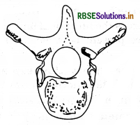
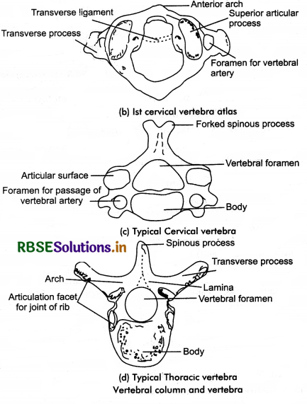
Identify the bone and comment on it.
Answer:
It is a typical thoracic vertebra it form the part of vertebral column there are 12 such vertebrae.
Question 24.
Define true ribs.
Answer:
The first 7 ribs of human skeleton are called true ribs.
Question 25.
Mention the components of appendicular skeleton.
Answer:
Component of appendicular skeleton are:
- Limb bones (30 bones)
- Pectoral and pelvic girdles.
Short Answer Type Questions
Question 1.
Write in brief about amoeboid movement in human body.
Answer:
Amoeboid Movement: The streaming of protoplasm forms finger like projections called pseudopodia, (pseudo =false, podia =feet). The cells moves/locomotes in the direction of pseudopodium formed as in Amoeba. Cytoskeletal elements like microfilaments are also involved in amoeboid movement. These microfilaments are formed of myosin and actin proteins. Some specialised cells in one body like macrophages and leucocytes (WBCs) in blood (exhibit amoeboid movement), embryonic mesenchyma cells and other cells that move in between inter tissue spaces.
Question 2.
Write in short about movements in cells of human body.
Answer:
The cell movement is a complex phenomenon which is due to the actin network under the cell membrane. It has three components - Protrusion of the cellular edge, adhesion and de - adhesion of the cellular body and cytoskeleton contraction which helps in pulling the cell.
The cells of human body exhibit major three types of movements-
Muscular movement: It is exhibited by the muscle cells. It is caused by the contraction of the myofibrils of the muscle cell.
Amoeboid movement: It is exhibited by the leucocytes of the blood. It is involved in phagocytosis i.e. engulfing of the foreign particles and digesting them by the help of digestive enzymes. It is a kind of defence mechanism.
Ciliary movement: It takes place in tubular organs which are lined by ciliated epithelium. The cilia in the reproductive tract of females help in movement of eggs.
Question 3.
Classify the types of muscles on the basis of their location in human body. Write about any one type in brief.
Answer:
Classification of muscles is done on the basis of different criteria namely: (a) location, (b) appearance and (c) nature of regulation of their activities. On the basis of location of muscles in human body these are:
(i) Skeletal Muscle: Muscles attached/associated with bones, skeletal components of the body are called skeletal muscles. Skeletal muscles appear striped under the microscope. Hence, it is called striated muscles also. These muscles can be controlled by will, nervous system, i. e., the movement in them can be voluntarily done. Hence, these are also called voluntary muscles. These muscles primarily helps in change of body postures and locomotion.
Other names of skeletal muscles: Striped muscles, voluntary muscles, fast muscles and striated muscles.
(ii) Visceral Muscles: The hollow visceral organs in our body have visceral muscles in the innerwalls of the organs. These muscles do not show any striation and are smooth in appearance. These muscle cells are spindle shaped with a single large nucleus in centre. The sarcoplasm has parallely arranged myofilaments. These muscles are not under the control of our will (nervous system) i.e., involuntary.
Hence, these are also called involuntary muscles. When they contract they reduces the length of the tract and creates a wave like motion and helps in transportation of contents. Such as food in digestive tract and gametes in the genital tract. Other names of visceral muscles are smooth muscles, slow muscles, involuntary muscles and non - striated muscles.
(iii) Cardiac Muscles: The muscles found/present in heart wall are called cardiac muscles. Many cardiac muscle cells arrange themselves in branching pattern to
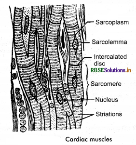
form a cardiac muscle. Cardiac muscles appear branched, striated, involuntary in nature as the nervous system does not control their activities directly. They possess the quality or both striated and smooth muscles. The connective tissue is loosely present in between the fibres which is transported by blood cells. Sarcolemma is not clearly distinct but one/single nucleus is present in centre. Myofibrils I - bands, A - bands and Z - line is clearly visible. At frequent intervals there are discs called intercalated disc between the I - bands and A - bands. Due to which the fibres are divisible in small compartments. The contraction in cardiac muscle fibres is sequential and they do not get tired.
Question 4.
Justify the reason for banded or striated appearance of skeletal muscle.
Answer:
Muscle is a specialised tissue originated from the germ layer, mesoderm. The muscular tissue is made up of specialised cells called myoctes. These cells are bounded together by a connective tissue and form muscular tissue.
Structure of Muscle: A muscle is covered by a sheath of connective tissue called epimysium. Inside the epimysium, a muscle has many muscle fibres arranged in a bundle called fasciculi. Each fasciculus surrounded by a sheath of connective called perimysium. The muscle fibres are parallel to each other in fasciculus and is surrounded by a connective tissue called endomysium. The muscle bundle are further bounded together by a common collagenous sheath of connective tissue called fascia.
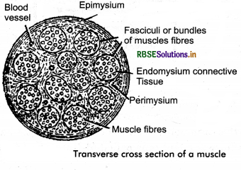
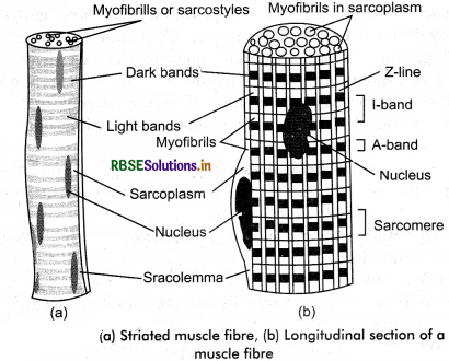
Let us study the detailed structure of striated muscle. It helps us to understand the mechanism of contraction of muscles. Each organised striated/skeletal muscle is made of a number of muscle bundles or fascicles held together by a common collagenous connective tissue layer called fascia. Each muscle bundle consists of many muscle fibres and each muscle fibre having a plasma membrane is called sarcolemma. the cytoplasm is called scarcoplasm. There are many nuclei present in the sarcoplasm of the muscle fibre. This situation of cell is called syncytium. The endoplasmic reticulum (ER) is called sarcoplasmic reticulum which are the store house of calcium ions.
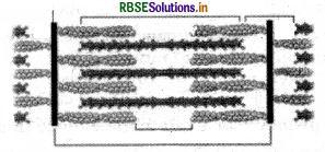
Muscle fibres have large number of parallely arranged filaments called myofilaments or myofibrils in sarcoplasm. Each myofilament/myofibril is made up of light and dark bands arranged alternately. These bands give a striated/striped appearance due to their arrangement in the muscle fibre. Two proteins actin and myosin form their bands. Light band is made of actin protein and it is called I - band or isotropic band. Dark band is made of myosin protein and it is called ‘A’ - band or Anisotropic band. Both of these proteins are arranged as rod - like structures which are parallel to each other and also, to the longitudinal axis of the myofibrils.

Actin filaments are thinner than the myosin filaments and hence, these are also termed as thin and thick filaments respectively. In the centre of each I - band is an elastic fibre called ‘Z’ - line which bisects it. Thin filaments are firmly attached to it.

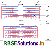
A - bands are arranged alternately throughout the length of the myofibrils. The portion of the myofibril between two successive ‘Z’ - lines is considered as the functional unit of the contraction and is called a sarcomere. The edges of the thin filaments on either side of the thick filaments partially overlap the free ends or the thick filaments leaving the central part of the thick filament (without overlap). This un - overlapped portion of the thick filaments forms a central part called ‘H’- zone. This condition is observed during the resting state of the muscle fibre.
Question 5.
Label the above figure and write in brief about sarcomere.
Answer:
Let us study the detailed structure of striated muscle. It helps us to understand the mechanism of contraction of muscles. Each organised striated/skeletal muscle is made of a number of muscle bundles or fascicles held together by a common collagenous connective tissue layer called fascia. Each muscle bundle consists of many muscle fibres and each muscle fibre having a plasma membrane is called sarcolemma. the cytoplasm is called scarcoplasm. There are many nuclei present in the sarcoplasm of the muscle fibre. This situation of cell is called syncytium. The endoplasmic reticulum (ER) is called sarcoplasmic reticulum which are the store house of calcium ions.
Muscle fibres have large number of parallely arranged filaments called myofilaments or myofibrils in sarcoplasm. Each myofilament/myofibril is made up of light and dark bands arranged alternately. These bands give a striated/striped appearance due to their arrangement in the muscle fibre. Two proteins actin and myosin form their bands. Light band is made of actin protein and it is called I - band or isotropic band. Dark band is made of myosin protein and it is called ‘A’ - band or Anisotropic band. Both of these proteins are arranged as rod - like structures which are parallel to each other and also, to the longitudinal axis of the myofibrils.

Actin filaments are thinner than the myosin filaments and hence, these are also termed as thin and thick filaments respectively. In the centre of each I - band is an elastic fibre called ‘Z’ - line which bisects it. Thin filaments are firmly attached to it.

A - bands are arranged alternately throughout the length of the myofibrils. The portion of the myofibril between two successive ‘Z’ - lines is considered as the functional unit of the contraction and is called a sarcomere. The edges of the thin filaments on either side of the thick filaments partially overlap the free ends or the thick filaments leaving the central part of the thick filament (without overlap). This un - overlapped portion of the thick filaments forms a central part called ‘H’- zone. This condition is observed during the resting state of the muscle fibre.
Question 6.
(i) What are contractile proteins?
(ii) Draw a labelled diagram of contractile proteins.
Answer:
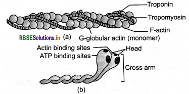
(i) The proteins that helps in formation of thick and thin filaments and which helps in muscle contraction are called contractile proteins.
(ii)
Question 7.
Give the diagrammatic representation of sliding filament theory of muscle contraction.
Answer:
Question 8.
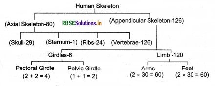
Classify (the division ) on human skeleton. Also, mention the number of bones.
Answer:
Question 9.
Write the characteristics of skull or vertebral column.
Answer:
Joints are the points of contact between bones or between bones and cartilage. They are essential for all types of movements involving the body parts of the body and of course, the locomotion too. The force created by the muscles is used to carry out the movement through joints where they acts as fulcrum. Various factors are responsible for the movements of these joints. Structurally the joints have been classified into 3 types:
1. Fibrous joints: The joints between the bones with the help of dense fibrous connective tissues are called fibrous joints. They do not allow any movement and are immovable (joints). Such as the fact skull bones fuse end to end with dense fibro connective tissue in the form of structures, joint between teeth and maxilla.
2. Cartilaginous joints: These joints are joined with the help of cartilages. Such as the joints between the adjacent vertebrae in the vertebral column. They show limited movements.
3. Synovial joints: There is presence of a fluid known as synovial fluid between the two articulating surfaces of the two bones in the synovial cavity. These joints have free movement in all the directions smoothly. These joints helps in movement and locomotion. They are called movable joints and categorised as:
(a) Pivot joint: Between atlas and axis. One bone forms pivot and other is axis.
(b) Saddle joint: Between carpal and metacarpal.
(c) Gliding joint: Between the carpal. The joints have flat bones that slide over each other: e.g., wrist joint.
(d) Hinge joint: This is between humerus and radio ulna the elbow joint and femur and tibia fibula in the knee joint. They move in one direction only i.e., folding and opening like a door or window that can be opened/closed.

(c) Ball and Socket joint: Between the humer head and glenoid cavity, femur head and acetabulum cavity. The head is in ball and socket is the depression in the cavity. Movement is in all direction 360°.
The joint between the pubic bones is pubic symphysis it is slightly movable.
Disorders of Muscular and Skeletal System
- Myasthenia gravis: A disorder of auto immune disorder which affects the neuromuscular junction leading to fatigue, weakening and paralysis of the skeletel muscles.
- Muscular dystrophy: Due to genetic disorder there is progressive degeneration of muscles.
- Tetany: Due to low Ca++ in body rapid muscular spasms (wild contractions) is called tetany.
- Arthritis: Inflammation of joints.
- Osteoporosis: The condition of decreased bone mass the bone becomes prone to fractures. It is age related disorder. Decreased levels of estrogen is the common cause.
- Gout: Due to accumulation of uric acid crystals in joints inflammation takes place at joints. It is called gout.
- Sprain: The tearing off or stretching of tendons and ligaments is called sprain. The portion is identified by inflammation and pain.
Question 10.
Write about the different types of ribs present in our body.
Answer:
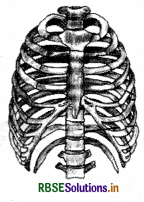
Stremum and ribs cage: Sternum is a flat bone on the ventral midline of thorax. It has 7 flat bones. These are divisible into three groups: presternum, mesosternum, xiphoid sternum. The 7 bones are collectively called sternebrae to which first 7 pairs of ribs are attached.
Ribs: There are 12 pairs of ribs. Each rib is a flat bone which is connected ventrally to the strenum and dorsally to vertebral column. It has two articulation surfaces on its dorsal end and is hence called bicephalic. Ribs are:
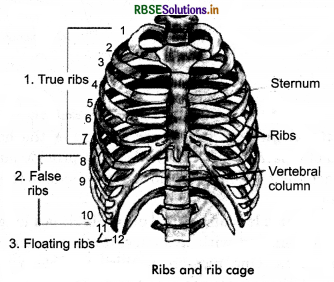
- True ribs: The first 7 ribs are called as true ribs. With the help of hyaline cartilage these are attached ventrally to sternum and at dorsal part to vertebral column.
- False ribs : The 8th, 9th and 10th pairs of ribs do not directly attached to sternum but they join to 7th rib with the help of hyaline cartilage. These are called vertebrochondral or false ribs.
- Floating ribs : The 11th and 12th pairs of last ribs are not connected ventrally and are free. Hence, these are called floating ribs.
Thoracic vertebrae, ribs and strenum are together (collectively) form the cage like structure called rib cage. Inside the rib cage is pair of lungs and heart.
Question 11.
Draw the diagram of following bones and label it.
(a) Humerus
(b) Radius, ulna
(c) Femur
(d) Tibia fibula
Answer:
Question 12.
Briefly write about fibrous and cartilagenous joints.
Answer:
1. Fibrous joints: The joints between the bones with the help of dense fibrous connective tissues are called fibrous joints. They do not allow any movement and are immovable (joints). Such as the fact skull bones fuse end to end with dense fibro connective tissue in the form of structures, joint between teeth and maxilla.
2. Cartilaginous joints: These joints are joined with the help of cartilages. Such as the joints between the adjacent vertebrae in the vertebral column. They show limited movements.
Question 13.
Label the figure: True ribs, false ribs, floating ribs, sternum, vertebral column. Write about the ribs.
Answer:
Stremum and ribs cage: Sternum is a flat bone on the ventral midline of thorax. It has 7 flat bones. These are divisible into three groups: presternum, mesosternum, xiphoid sternum. The 7 bones are collectively called sternebrae to which first 7 pairs of ribs are attached.
Ribs: There are 12 pairs of ribs. Each rib is a flat bone which is connected ventrally to the strenum and dorsally to vertebral column. It has two articulation surfaces on its dorsal end and is hence called bicephalic. Ribs are:

- True ribs: The first 7 ribs are called as true ribs. With the help of hyaline cartilage these are attached ventrally to sternum and at dorsal part to vertebral column.
- False ribs : The 8th, 9th and 10th pairs of ribs do not directly attached to sternum but they join to 7th rib with the help of hyaline cartilage. These are called vertebrochondral or false ribs.
- Floating ribs : The 11th and 12th pairs of last ribs are not connected ventrally and are free. Hence, these are called floating ribs.
Thoracic vertebrae, ribs and strenum are together (collectively) form the cage like structure called rib cage. Inside the rib cage is pair of lungs and heart.
Question 14.
Write in brief about the following:
(i) Muscular dystrophy.
(ii) Osteoporosis.
(iii) Sprain.
Answer:
(i) Muscular dystrophy: Due to genetic disorder there is progressive degeneration of muscles.
(ii) Osteoporosis: The condition of decreased bone mass the bone becomes prone to fractures. It is age related disorder. Decreased levels of estrogen is the common cause.
(iii) Sprain: The tearing off or stretching of tendons and ligaments is called sprain. The portion is identified by inflammation and pain.
Question 15.
Mention in short about any three synovial joints.
Answer:
Synovial joints: There is presence of a fluid known as synovial fluid between the two articulating surfaces of the two bones in the synovial cavity. These joints have free movement in all the directions smoothly. These joints helps in movement and locomotion. They are called movable joints and categorised as:
- Pivot joint: Between atlas and axis. One bone forms pivot and other is axis.
- Saddle joint: Between carpal and metacarpal.
- Gliding joint: Between the carpal. The joints have flat bones that slide over each other: e.g., wrist joint.
- Hinge joint: This is between humerus and radio ulna the elbow joint and femur and tibia fibula in the knee joint. They move in one direction only i.e., folding and opening like a door or window that can be opened/closed.
Long Answer Type Questions
Question 1.
Briefly explain the types of movements in animals.
Answer:
The human body cells show three main types of movements: amoeboid, ciliary and muscular.
1. Movement by Cellular Organelles: The cellular organelles like cilia and flagella are the locomotory organs in ciliates and flagellates. In human body cells, amoeboid and ciliary movements are also found.
(a) Amoeboid Movement: The streaming of protoplasm forms finger like projections called pseudopodia, (pseudo =false, podia =feet). The cells moves/locomotes in the direction of pseudopodium formed as in Amoeba. Cytoskeletal elements like microfilaments are also involved in amoeboid movement. These microfilaments are formed of myosin and actin proteins. Some specialised cells in one body like macrophages and leucocytes (WBCs) in blood (exhibit amoeboid movement), embryonic mesenchyma cells and other cells that move in between inter tissue spaces.
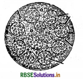
(b) Ciliary Movement: The movements caused due to the beating cilia (sing.cilium) is called ciliary movement. Cilia are small hair like ectoplasmic projections of the cell. They are very mobile help in movement of food in cytopharynx in ciliates like: Paramoecium and helps in locomotion.
In our body ciliary movement occurs in nose or our internal tubular organs. These organs are lined with ciliated Epithelium. The movement of materials in these internal organs is because of co - ordinated movements of cilia.
(c) Flagellar Movement: Movements caused due to beating/strokes of flagellum (pi.flagella) are called flagellar movement. It is also called paddle movement. Flagellum is a long whip like structure, the outgrowth of the cell membrane. This movement helps in swimming of spermatozoa, maintenance of water current in the water canal system of sponges and locomotion in protozoan: Euglena.
2. Movement of body Appendages: Many animals have appendages that helps the animal in movement and locomotion. Such as in invertebrates:
- Setae and parapodia: earthworms and other annelids.
- Tentacles: in Hydra, Physelia.
- Jointed appendages in insects and crustaceans: crabs.
- Tube feet: in echinoderms and starfish.
- Fins: in fishes.
- Limbs: fore arms and hind limbs in vertebrates.
3. Movements by Muscles: Muscles are specialised tissue of mesodermal origin. The human body weight (about 40 - 50 percent) is due to muscles. They have special properties like excitibility, elasticity, extensibility and contractibility. The movements due to muscles are called muscular movements.
Question 2.
Describe the structure of skeletal muscles.
Answer:
Let us study the detailed structure of striated muscle. It helps us to understand the mechanism of contraction of muscles. Each organised striated/skeletal muscle is made of a number of muscle bundles or fascicles held together by a common collagenous connective tissue layer called fascia. Each muscle bundle consists of many muscle fibres and each muscle fibre having a plasma membrane is called sarcolemma. the cytoplasm is called scarcoplasm. There are many nuclei present in the sarcoplasm of the muscle fibre. This situation of cell is called syncytium. The endoplasmic reticulum (ER) is called sarcoplasmic reticulum which are the store house of calcium ions.
Muscle fibres have large number of parallely arranged filaments called myofilaments or myofibrils in sarcoplasm. Each myofilament/myofibril is made up of light and dark bands arranged alternately. These bands give a striated/striped appearance due to their arrangement in the muscle fibre. Two proteins actin and myosin form their bands. Light band is made of actin protein and it is called I - band or isotropic band. Dark band is made of myosin protein and it is called ‘A’ - band or Anisotropic band. Both of these proteins are arranged as rod - like structures which are parallel to each other and also, to the longitudinal axis of the myofibrils.

Actin filaments are thinner than the myosin filaments and hence, these are also termed as thin and thick filaments respectively. In the centre of each I - band is an elastic fibre called ‘Z’ - line which bisects it. Thin filaments are firmly attached to it.

A - bands are arranged alternately throughout the length of the myofibrils. The portion of the myofibril between two successive ‘Z’ - lines is considered as the functional unit of the contraction and is called a sarcomere. The edges of the thin filaments on either side of the thick filaments partially overlap the free ends or the thick filaments leaving the central part of the thick filament (without overlap). This un - overlapped portion of the thick filaments forms a central part called ‘H’- zone. This condition is observed during the resting state of the muscle fibre.
Question 3.
Draw the diagram of sarcomere and explain its structure.
Answer:
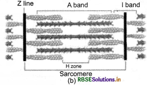
The thick filaments are also held together in A - band by a thin fibrous membrane called M - line. The I - bands and A - bands are arranged alternately throughout the length of the myofibrils. The portion of the myofibril between two successive ‘Z’ - lines is considered as the functional unit of the contraction and is called a sarcomere.
Question 4.
Explain about actin filament and myosin filament. Support your explanation with suitable diagrams.
Answer:
Let us study the detailed structure of striated muscle. It helps us to understand the mechanism of contraction of muscles. Each organised striated/skeletal muscle is made of a number of muscle bundles or fascicles held together by a common collagenous connective tissue layer called fascia. Each muscle bundle consists of many muscle fibres and each muscle fibre having a plasma membrane is called sarcolemma. the cytoplasm is called scarcoplasm. There are many nuclei present in the sarcoplasm of the muscle fibre. This situation of cell is called syncytium. The endoplasmic reticulum (ER) is called sarcoplasmic reticulum which are the store house of calcium ions.
Muscle fibres have large number of parallely arranged filaments called myofilaments or myofibrils in sarcoplasm. Each myofilament/myofibril is made up of light and dark bands arranged alternately. These bands give a striated/striped appearance due to their arrangement in the muscle fibre. Two proteins actin and myosin form their bands. Light band is made of actin protein and it is called I - band or isotropic band. Dark band is made of myosin protein and it is called ‘A’ - band or Anisotropic band. Both of these proteins are arranged as rod - like structures which are parallel to each other and also, to the longitudinal axis of the myofibrils.
Actin filaments are thinner than the myosin filaments and hence, these are also termed as thin and thick filaments respectively. In the centre of each I - band is an elastic fibre called ‘Z’ - line which bisects it. Thin filaments are firmly attached to it.
A - bands are arranged alternately throughout the length of the myofibrils. The portion of the myofibril between two successive ‘Z’ - lines is considered as the functional unit of the contraction and is called a sarcomere. The edges of the thin filaments on either side of the thick filaments partially overlap the free ends or the thick filaments leaving the central part of the thick filament (without overlap). This un - overlapped portion of the thick filaments forms a central part called ‘H’- zone. This condition is observed during the resting state of the muscle fibre.
Question 5.
Briefly explain each stages of muscle contraction and relaxation.
Answer:
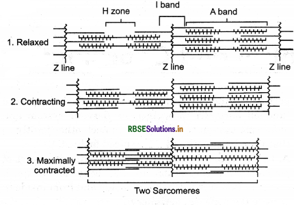
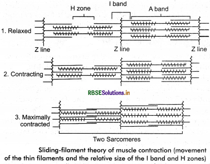
Sliding filament theory of muscle contraction was proposed by Huxley in 1965. According to it the contraction of a muscle fibre takes place by from both the sides of their filaments slide over the thick filaments.
Initiation of Muscle Contraction: The initiation of muscle contraction is signaled by the central nervous system (CNS) via motor nerve. Muscle fibres alongwith a motor nerve/nerves is connected to it forms a motor unit. The function between a motor nerve and the sarcolemma on the muscle fibre is neuromuscular junction or motor end plate.
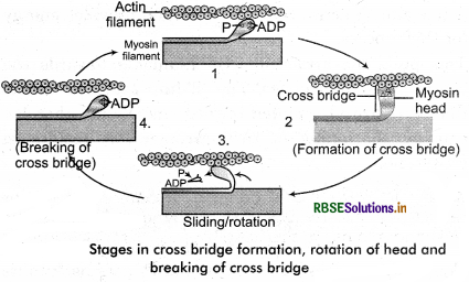
When the signal reaches the junction, a neuro transmitter is released. Acetyl choline is released (neurotransmitter) which creates an action potential in the sarcolemma. It spreads through the muscle fibre and reaches the release of calcium ions (Ca++) into the sarcoplasm. The increase in Ca++ level leads to the binding of calcium with a subunit of troponin on active filaments and thereby remove the masking of active sites of myosin protein (thick filament). Cross bridges are formed by the binding of myosin head to the exposed active site on actin. Hydrolysis of ATP provides energy for this process.
This pulls the attached actin filaments towards the centre of A - band (⇌). The ‘Z’ - line attached to the filaments are also pulled towards inside and thereby, causing shortening of the sarcomere, it results in contraction.
The myosin, releasing the ADP and Pi goes back to its relaxed state. A new ATP binds and the cross - bridge is broken. The cycle of cross bridge formation again is repeated by the hydrolysis of ATP by myosin head and breakage is repeated and causing further sliding. Till the Ca++ ions are pumped back causing to the sarcoplasmic cisternae results in masking of the actin filaments. The process continues. Relaxation of muscles is caused by the return of ‘Z’ - lines back to the original position.
Question 6.
Diagrammatically explain sliding filament theory proposed by Huxley.
Answer:
The sliding filament theory explains the mechanism of muscle contraction based on muscle proteins that slide past each other to generate movement. According to the sliding filament theory, the myosin (thick filaments) of muscle fibers slide past the actin (thin filaments) during muscle contraction, while the two groups of filaments remain at relatively constant length.
The theory was independently introduced in 1954 by two research teams, one consisting of Andrew Huxley and Rolf Niedergerke from the University of Cambridge, and the other consisting of Hugh Huxley and Jean Hanson from the Massachusetts Institute of Technology. It was originally conceived by Hugh Huxley in 1953. Andrew Huxley and Niedergerke introduced it as a "very attractive" hypothesis.
Before the 1950s there were several competing theories on muscle contraction, including electrical attraction, protein folding, and protein modification.
The novel theory directly introduced a new concept called cross - bridge theory (classically swinging cross - bridge, now mostly referred to as (cross - bridge cycle) which explains the molecular mechanism of sliding filament. Cross - bridge theory states that actin and myosin form a protein complex (classically called actomyosin) by attachment of myosin head on the actin filament, thereby forming a sort of cross - bridge between the two filaments. The sliding filament theory is a widely accepted explanation of the mechanism that underlies muscle contraction.
Question 7.
Briefly mention about axial skeleton.
Answer:
Axial skeleton: The axis of the body is formed of 80 bones. It is called axial skeleton. It consists of:
- Skull
- Vertebral column (back bone)
- Sternum and ribs (rib cage).
(a) Skull: Human skull is composed of two sets of bones: cranial and facial bones. There are 29 bones in skull. Cranial bones are 8 in number. They form a hard protective outer covering called cranium in which the brain is protected and lodged. Facial region or skull is formed of 14 skeletal elements, it forms the front part of the skull. A single 'U' shaped bone called hyoid is present at the base of the buccal cavity and it is also included in the skull. Each of the middle ear has 3 tiny bones called ear ossicles these are Malleus, Incus and Stapes (MIS). They have hammer, anvil and stapes (HAS) shape respectively.
The skull region articulates with the superior region of the vertebral column with the help of two occipital condyles. This type of skull is known as dicondvlic skull.
Characteristics of skull
- It is completely made up of bones.
- It is dicondylic.
- It has large/big cranial capacity.
- It has zygometic arch at posterior part.
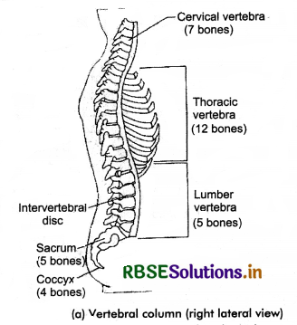
(b) Vertebral Column (back bone): The vertebral column is made up of 26 serially arranged units of irregular shape called vertebrae (sing, vertebra). It is also called back bone because of its location in the posterior part of the human body (i.e., dorsally placed). The vertebral column extends from the base of the skull and constitutes the main framework of the trunk. In each vertebra, is present a central hollow portion called neural canal the spinal cord passes through it.
First vertebra of the vertebral column is called atlas. It articulates with the occipital condyles of the skull. The vertebral column can be differentiated into (from skull):
(i) Cervical: consists of 7 vertebrae (bones).
(ii) Thoracic: consists of 12 vertebrae.
(iii) Lumbar: consists of 5 vertebrae.
(iv) Sacral: consists of 5 bones fused as 1 bone.
(v) Coccygeal: consists of 4 bones fused as 1 bone. Spinal cord (extension) of the brain passes through the neural canal of the vertebrae or the vertebral column. Thus, the vertebral column protects spinal cord, head and provides point to attachment for the ribs and musculature of the back.
Characteristics of Vertebral Column are:
- The centrum is of amphiplatyan or acoelus type.
- A fibrous disc is present in between two vertebrae. It provides it flexibility.
- On each point of strenum the plate of epiphysis of vertebrae are attached.
- The capacity of vertebral column’s flexibility/ bending’s due to the joint of anterior arch of one vertebra to the posterior part of the next vertebra.
- Spinal cord is protected by the vertebral column.
(c) Stremum and ribs cage: Sternum is a flat bone on the ventral midline of thorax. It has 7 flat bones. These are divisible into three groups: presternum, mesosternum, xiphoid sternum. The 7 bones are collectively called sternebrae to which first 7 pairs of ribs are attached.
Ribs: There are 12 pairs of ribs. Each rib is a flat bone which is connected ventrally to the strenum and dorsally to vertebral column. It has two articulation surfaces on its dorsal end and is hence called bicephalic. Ribs are:

- True ribs: The first 7 ribs are called as true ribs. With the help of hyaline cartilage these are attached ventrally to sternum and at dorsal part to vertebral column.
- False ribs : The 8th, 9th and 10th pairs of ribs do not directly attached to sternum but they join to 7th rib with the help of hyaline cartilage. These are called vertebrochondral or false ribs.
- Floating ribs : The 11th and 12th pairs of last ribs are not connected ventrally and are free. Hence, these are called floating ribs.
Thoracic vertebrae, ribs and strenum are together (collectively) form the cage like structure called rib cage. Inside the rib cage is pair of lungs and heart.
Question 8.
Mention in brief about fore limbs. Give a diagram also.
Answer:
(a) Limb bones: The limbs are in a pair.
Fore limbs and hind limbs
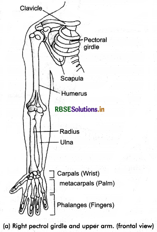
Fore limbs: These are also called fore limbs/arms/hands.
(i) It has upper arm madeup of a long bone called humerus. It is cylindrical, hollow bone filled with bone marrow in the long portion called shaft. The upper end has a ball that fits into the glenoid cavity of shoulder bone and the lower end has two ball like bones that fits into the socket of fore arm.
(ii) The fore arm is formed of two long bones: Radius and ulna, radius is towards the outside and ulna is towards the body, i.e., the bone towards thumb is radius and the bone towards little finger is ulna. There is a gap in between the bones from top to bottom.
(iii) Hand/Palm/Manus is the broad part of the fore limb, it has:
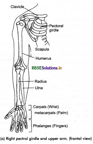
(a) Wrist: It is made of 8 bones, arranged as 4 - 4 in 2 rows. These are called carpals. These are flexible angle they can be moved in any direction.
(b) Palm is formed of 5 bones they are long metacarpals.
(c) Fingers are the phalanges, the digits are 14 in number. They are attached to each other. In a single finger 3 bones are present and 2 bones in thumb. Thus, all the 5 fingers have 14 phalanges.
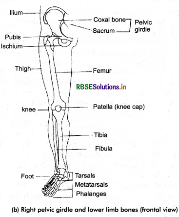
Hind limbs are also called legs. Both the legs/hind limbs in total have 60 bones.
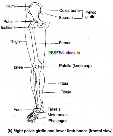
(i) The upper portion of the leg is called thigh. It has the longest hone of the body called femur. It has a head with a ball like structure that fits into the acetubulum cavity or the pelvic girdle. The mid part is called shaft and is hollow with bone marrow packed in it. The other end of the bone has 2 ball likes structures that fit into the depressions on tibia and fibula. This joint is covered with a round triangular cup called knee cap (patella). It helps to provides movement/speed and the knee can be folded.
(ii) The next part in legs formed of two bones having wide gap in between them. They are separate. Tibia is a comparatively thick and located inside towards the body and fibula is thin, weak and located on outer side.
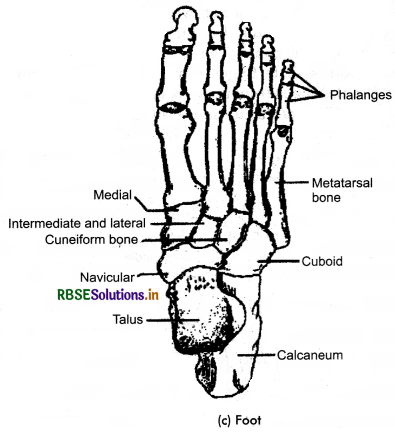
(iii) Foot: It is the basal portion of body. It helps in standing body.
(a) Ankle is formed of 7 small bones attached to tibia and fibula. These are called as tarsals. It forms ankle and heel.
(b) Sole foot is formed of 5 long bones called metatarsals.
(c) Fingers are made of phalanges joined to metatarsals. Each finger has 3 bones and thumb has 2 bones. In total 14 bones are present.
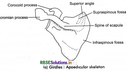
(a) Pectoral girdle: The upper portion of the body is formed of a pair of pectoral girdles that provide support for attachment to the fore limb on either side to the axial skeleton.
(i) Collar bone: It is also called clavicle. It is a long bone slightly 'S’ shaped with two curvatures, present above the first ribs. One portion of this is joint in mid to sternum. Other portion is broad and flat and it is joined to scapula.
(ii) Scapula: It is a broad, flat and triangular bone. It is located below the shoulder and the back portion. At the end of the mid line, a projection/process called acromian process is present, (i.e., dorsal part of the thorax between the 2nd and 7th ribs.) It has a slightly elevated ridge called spine which the outer portion the clavicle/collar bone is attached to it. Below this processus present the glenoid cavity into which the head of humerus fits.
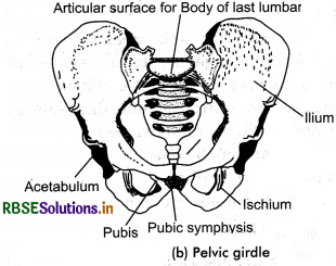
(b) Pelvic girdle: This girdle is also called hip girdle. This girdle is formed of two coxal bones joint together (with sacrum) to provide support to sacrum in pelvic region.
It is also called hip bones. Towards the another part these are attached to pubic symphysis containing fibrous cartilage and to posterior jointed to sacrum. Each coxal bone is formed of (i) ilium, (ii) pubis and
(iii) ischium. Which laber these to form the girdle. At the junction of these three bones in a depression called acetabulum to which the head of the femur fits in on the either sides.
Functions of the Endoskeleton
- It provides the body framework.
- It protects the delicate internal organs of the body.
- Along with muscles provides locomotion.
- It provides a surface for attachment to muscles.
- It makes straight/erect posture of the human body.
- It has redbone marrow in hollow bones manufacture RBCs (erythropoiesis).
- Ear ossicles helps in hearing process.
- Thoracic cavity helps in respiration process.
- Bone marrow is the store house for fats, minerals such as: Ca and PO4.
Question 9.
Write about different types of movable joints present in your body with one example to each.
Answer:
Joints are the points of contact between bones or between bones and cartilage. They are essential for all types of movements involving the body parts of the body and of course, the locomotion too. The force created by the muscles is used to carry out the movement through joints where they acts as fulcrum. Various factors are responsible for the movements of these joints. Structurally the joints have been classified into 3 types:
1. Fibrous joints: The joints between the bones with the help of dense fibrous connective tissues are called fibrous joints. They do not allow any movement and are immovable (joints). Such as the fact skull bones fuse end to end with dense fibro connective tissue in the form of structures, joint between teeth and maxilla.
2. Cartilaginous joints: These joints are joined with the help of cartilages. Such as the joints between the adjacent vertebrae in the vertebral column. They show limited movements.
3. Synovial joints: There is presence of a fluid known as synovial fluid between the two articulating surfaces of the two bones in the synovial cavity. These joints have free movement in all the directions smoothly. These joints helps in movement and locomotion. They are called movable joints and categorised as:
(a) Pivot joint: Between atlas and axis. One bone forms pivot and other is axis.
(b) Saddle joint: Between carpal and metacarpal.
(c) Gliding joint: Between the carpal. The joints have flat bones that slide over each other: e.g., wrist joint.
(d) Hinge joint: This is between humerus and radio ulna the elbow joint and femur and tibia fibula in the knee joint. They move in one direction only i.e., folding and opening like a door or window that can be opened/closed.
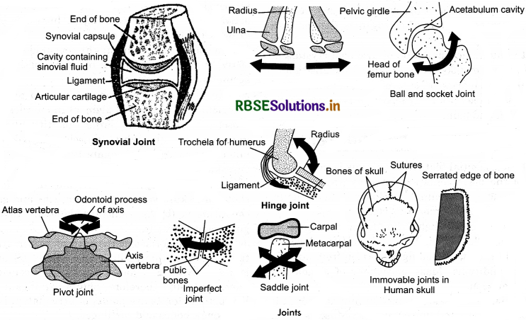
(c) Ball and Socket joint: Between the humer head and glenoid cavity, femur head and acetabulum cavity. The head is in ball and socket is the depression in the cavity. Movement is in all direction 360°.
The joint between the pubic bones is pubic symphysis it is slightly movable.
Disorders of Muscular and Skeletal System
- Myasthenia gravis: A disorder of auto immune disorder which affects the neuromuscular junction leading to fatigue, weakening and paralysis of the skeletel muscles.
- Muscular dystrophy: Due to genetic disorder there is progressive degeneration of muscles.
- Tetany: Due to low Ca++ in body rapid muscular spasms (wild contractions) is called tetany.
- Arthritis: Inflammation of joints.
- Osteoporosis: The condition of decreased bone mass the bone becomes prone to fractures. It is age related disorder. Decreased levels of estrogen is the common cause.
- Gout: Due to accumulation of uric acid crystals in joints inflammation takes place at joints. It is called gout.
- Sprain: The tearing off or stretching of tendons and ligaments is called sprain. The portion is identified by inflammation and pain.

- RBSE Solutions for Class 11 Biology Chapter 10 Cell Cycle and Cell Division
- RBSE Solutions for Class 11 Biology Chapter 9 Biomolecules
- RBSE Solutions for Class 11 Biology Chapter 8 Cell: The Unit of Life
- RBSE Solutions for Class 11 Biology Chapter 7 Structural Organisation in Animals
- RBSE Solutions for Class 11 Biology Chapter 6 Anatomy of Flowering Plants
- RBSE Solutions for Class 11 Biology Chapter 5 Morphology of Flowering Plants
- RBSE Solutions for Class 11 Biology Chapter 4 Animal Kingdom
- RBSE Solutions for Class 11 Biology Chapter 3 Plant Kingdom
- RBSE Solutions for Class 11 Biology Chapter 2 Biological Classification
- RBSE Solutions for Class 11 Biology Chapter 1 The Living World
- RBSE Solutions for Class 11 Biology Chapter 5 पुष्पी पादपों की आकारिकी