RBSE Class 11 Biology Important Questions Chapter 18 Body Fluids and Circulation
Rajasthan Board RBSE Class 11 Biology Important Questions Chapter 18 Body Fluids and Circulation Important Questions and Answers.
Rajasthan Board RBSE Solutions for Class 11 Biology in Hindi Medium & English Medium are part of RBSE Solutions for Class 11. Students can also read RBSE Class 11 Biology Important Questions for exam preparation. Students can also go through RBSE Class 11 Biology Notes to understand and remember the concepts easily.
RBSE Class 11 Biology Chapter 18 Important Questions Body Fluids and Circulation
Multiple Choice Questions
Question 1.
Blood circulation was discovered by:
(a) William Watson
(b) William Harvey
(c) Landsteiner
(d) P. Venugopal
Answer:
(b) William Harvey

Question 2.
The heart of human is:
(a) Myogenic
(b) Neurogenic
(c) Partialy myogenic
(d) Partialy neurogenic
Answer:
(a) Myogenic
Question 3.
Venous heart is found in:
(a) Fishes
(b) Frog
(c) Crocodile
(d) Rabbit
Answer:
(a) Fishes
Question 4.
The contraction in heart starts from:
(a) Right atrium
(b) Right ventricle
(c) Left atrium
(d) Left ventricle
Answer:
(a) Right atrium
Question 5.
Ventricular contraction is controlled by:
(a) SAN
(b) AVN
(c) Purkinje fibres
(d) Papillary muscles
Answer:
(a) SAN
Question 6.
Sino - auricular node is present in heart is:
(a) Right ventricle
(b) Left ventricle
(c) Right atrium
(d) Left atrium
Answer:
(c) Right atrium
Question 7.
Haematology refers to the study of:
(a) Bones
(b) Blood
(c) Cartilage
(d) Nerves
Answer:
(b) Blood
Question 8.
Atheroma is related with:
(a) Blood vessels
(b) Ureter
(c) Trachea
(d) Hepatic duct
Answer:
(a) Blood vessels
Question 9.
Primary blood cells are formed in:
(a) Plasma
(b) Bone marrow
(c) Spleen
(d) Liver
Answer:
(b) Bone marrow
Question 10.
The wall of heart is made of:
(a) Epicardium
(b) Myocardium
(c) Endocardium
(d) All of these
Answer:
(d) All of these
Question 11.
Person having blood group A will having:
(a) A antigen and B antibody
(b) B antigen and A antibody
(c) Both A and B antigens
(d) Antigen and antibodies absent
Answer:
(a) A antigen and B antibody
Question 12.
Slowing down of rate of heart beat is called:
(a) Arrythemia
(b) Trachycardia
(c) Bradycardia
(d) All of these
Answer:
(c) Bradycardia
Question 13.
Neurogenic heart is found in:
(a) Cockroach
(b) Humans
(c) Rat
(d) Rabbit
Answer:
(a) Cockroach
Question 14.
Universal donor is:
(a) O Rh+
(b) O Rh-
(c) AB Rh+
(d) AB Rh-
Answer:
(b) O Rh-
Question 15.
Universal recipient is:
(a) O Rh-
(b) O Rh+
(c) AB Rh+
(d) AB Rh-
Answer:
(c) AB Rh+
Question 16.
What is found in antiserum?
(a) Antigens
(b) Antibodies
(c) WBCs
(d) None of these
Answer:
(b) Antibodies
Question 17.
Purkinje fibres are found in:
(a) Brain
(b) Blood
(c) Lungs
(d) Heart
Answer:
(d) Heart
Question 18.
Plasma protein which maintains the osmotic pressure of blood is:
(a) Albumins
(b) Globulins
(c) Prothrombin
(d) Fibrinogen
Answer:
(a) Albumins
Question 19.
Which protein helps in clot formation?
(a) Globulin
(b) Albumin
(c) Fibrinogen
(d) None of these
Answer:
(c) Fibrinogen
Question 20.
Pacemaker is situated in heart:
(a) In the wall of right atrium
(b) On inter articular septum
(c) Both A and B antigens
(d) Antigen and antibodies absent
Answer:
(a) In the wall of right atrium
Question 21.
Right atrium receives blood from:
(a) Pulmonary aorta
(b) Pulmonary veins
(c) Inferior venacava
(d) Superior and inferior vena cava
Answer:
(d) Superior and inferior vena cava
Question 22.
First heart sound is:
(a) ‘lubb’ due to closure of AV valves
(b) ‘lubb’ due to closure of spiral valves
(c) ‘dub’ due to closure of AV valves
(d) ‘dub’ due to closure of spiral valves
Answer:
(a) ‘lubb’ due to closure of AV valves
Question 23.
If vagus nerve to heart is cut, the heart beat will:
(a) decrease
(b) increase
(c) remain normal
(d) stop
Answer:
(b) increase
Question 24.
Hardening of the wall of small arteries is known as:
(a) Thrombosis
(b) Arteriosclerosis
(c) Atherosclerosis
(d) Myocardial infarction
Answer:
(b) Arteriosclerosis
Question 25.
The heart murmur due to:
(a) coronary thrombosis
(b) defective leaky valve
(c) arterial pulse
(d) all developed atrium
Answer:
(b) defective leaky valve
Question 26.
What is the stroke volume of an adult human heart?
(a) 50 mL
(b) 70 mL
(c) 90 mL
(d) 100 mL
Answer:
(b) 70 mL
Question 27.
Ventricular diastole occurs due to a/an:
(a) organ system
(b) cell organelle
(c) tissue
(d) organ
Answer:
(c) tissue
Question 28.
Cardiac muscle is found in:
(a) myocardium
(b) epicardium
(c) endocardium
(d) all of these
Answer:
(a) myocardium
Question 29.
Pulmonary vein carries:
(a) deoxygenated blood
(b) oxygenated blood
(c) mixed blood
(d) none of these
Answer:
(b) oxygenated blood
Question 30.
Heart of heart is:
(a) SA node
(b) AV node
(c) Bundle of His
(d) Purkinje fibres
Answer:
(a) SA node
Question 31.
Given below is the ECG of a normal human. Which one of its components is correctly interpreted below:
(a) Complex QRS - one complete pulse
(b) Peak T - initiation of total cardiac contraction
(c) Peak P and Peak R - together - systolic and diastolic blood pressure
(d) Peak P - initiation of left atrial contraction only
Answer:
(a) Complex QRS - one complete pulse
Question 32.
The cardiac cycle is normal subject is about:
(a) 0.5 second
(b) 0.8 second
(c) 1.0 second
(d) 1.2 second
Answer:
(b) 0.8 second
Question 33.
Chordae tendinae are found in:
(a) Atria of heart
(b) ventricle of heart
(c) joints of legs
(d) ventricles of brain
Answer:
(b) 0.8 second
Question 34.
Which one is help in blood clotting?
(a) Ca++
(b) Mg++
(c) K+
(d) Na+
Answer:
(a) Ca++
Question 35.
Erythrocytes are formed in:
(a) Thymus
(b) Liver
(c) Bone marrow
(d) Spleen
Answer:
(c) Bone marrow
Question 36.
Renal portal system is found in:
(a) All vertebrates
(b) All chordates
(c) Absent in mammals
(d) All mammals
Answer:
(c) Absent in mammals
Question 37.
The most found WBCs in blood are:
(a) Monocytes
(b) Basophils
(c) Neutrophils
(d) Eosinophils
Answer:
(c) Neutrophils
Question 38.
Rh factor is present in:
(a) All vertebrates
(b) All mammals
(c) All reptiles
(d) Human and Rhesus monkey
Answer:
(d) Human and Rhesus monkey
Question 39.
Which chamber of the human heart has the thickest muscular wall?
(a) Left auricle
(b) Left ventricle
(c) Right auricle
(d) Right ventricle
Answer:
(b) Left ventricle
Question 40.
Heart sound which is longer is:
(a) lubb
(b) dub
(c) both equal
(d) sometimes (a) and sometimes (b)
Answer:
(a) lubb
Very Short Answer Type Questions
Question 1.
Which type of human heart is?
Answer:
Human heart is myogenic type.
Question 2.
Where are chordae tendinae found?
Answer:
Chordae tendinae are found in ventricles. Their one end attached with ventricular wall and other one with atrio ventricular valve.
Question 3.
From where carotid systemic arch starts?
Answer:
Carotid systemic arch starts from left ventricle.
Question 4.
Where tricuspid valves are found?
Answer:
Tricuspid valves are found on right atrio - ventricular aperture.
Question 5.
Where bicuspid valve is present?
Answer:
Bicuspid valve is present on left atrio - ventricular aperture.
Question 6.
Where mitril valves are present?
Answer:
Bicuspid valve is called mitril valve it is present on left atrio - ventricular aperture.
Question 7.
Which type of blood flow in pulmonary vein?
Answer:
Pulmonary vein carries oxygenated blood.
Question 8.
Which parts of heart have pure and impure blood?
Answer:
Left part has pure blood and right part has impure blood.
Question 9.
Where pace makers situated in heart?
Answer:
Pacemaker are situated near eustachian valve in right auricle.
Question 10.
Name two pacemakers found in heart.
Answer:
- Sino - atrial node (SA node).
- Atrio - ventricular node (AV - node).
Question 11.
Name the two types of circulatory system.
Answer:
- Open circulatory system.
- Closed circulatory system.
Question 12.
Which type of circulatory system found in earth worm and cockroach?
Answer:
Earthworm - Closed circulatory system
Cockroach - Open circulatory system.
Question 13.
How many chambers are found in human heart? Name them.
Answer:
Four chambers: Two atria and two ventricles.
Question 14.
What is foramen ovalis?
Answer:
During foetal stage there is an aperture in the interatrial septum called foramen ovalis.
Question 15.
How blood supply in heart wall itself?
Answer:
Through coronary artery.
Question 16.
What is neurogenic heart?
Answer:
In this type of heart, the heart beat originates through nervous tissue.
Question 17.
What is heart beat?
Answer:
The regular and rhythmic contraction of heart is called heart beat.
Question 18.
What is systolic volume?
Answer:
The volume of blood pumped out by ventricle in one contraction is called systolic volume or stroke volume.
Question 19.
Name the device used in measuring blood pressure.
Answer:
Sphygmomanometer.
Question 20.
What is pulse?
Answer:
The rhythmic distension of the wall of the artery due to systolic rise in blood pressure is called pulse.
Question 21.
What is the pulse rate in an adult human and children?
Answer:
In an adult human: 70 - 90 per minute.
In children: 80 - 140 per minute.
Question 22.
Who called the father of electrocardiography?
Answer:
Einthoven called as father of electrocardiography.
Question 23.
Write the two main components of blood and their percentage.
Answer:
- Blood plasma: 55%.
- Blood corpuscles: 45%.
Question 24.
Which organ is called grave yard of RBCs?
Answer:
Spleen is known as grave yard of RBCs.
Question 25.
Who discovered blood groups?
Answer:
Carl Landsteiner (1900).
Question 26.
Who discovered Rh - factor?
Answer:
Landsteiner and Wiener.
Question 27.
Write the name of two main circuits of human circulatory system.
Answer:
- Pulmonary circulation.
- Systemic circulation.
Question 28.
Which protein is essential for blood clotting?
Answer:
Prothrombin protein is essential for blood clotting.
Question 29.
What are systole and diastole?
Answer:
Contraction of heart is called systole and relaxation of heart is called diastole.
Question 30.
Give one function of thrombocytes.
Answer:
They take part in blood clotting.
Question 31.
Give main function of lymphocytes.
Answer:
Lymphocytes produce antibodies.
Question 32.
Where antibodies are found?
Answer:
Antibodies are found in blood plasma.
Question 33.
What are antigens?
Answer:
The RBCs of human has specialized proteins aglutinogens, these are called antigens.
Question 34.
What is the base of classification of blood group?
Answer:
Antigen present on surface of RBCs and antibodies present in plasma.
Question 35.
What are constituents of lymph?
Answer:
Lymph consists of plasma and leucocytes.
Short Answer Type Questions - I
Question 1.
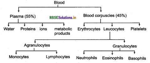
Give the diagrammatic representation of components of blood.
Answer:
Components of blood:
Question 2.
What is plasma? Name its component.
Answer:
Plasma is pale coloured, slightly alkaline clear, transparent, fluid without corpuscles. It constitutes about 55% of the blood. It has water 90 - 91%, inorganic substances 1%, plasma proteins 7% and other organic substances 1%.
Question 3.
Mention the similarities between blood and lymph.
Answer:
Similarities between blood and lymph:
- Both have white blood corpuscles.
- Both contain glucose, amino acid, vitamins, salt, hormones and urea in equal amount.
- Lymph has fibrinogen like blood and can make clot as blood.
- Lymph has antibody and antitoxin as in blood.
Question 4.
What are erythropoiesis and erythrolysis?
Answer:
The process of the production of erythrocytes is called erythropoiesis and the organ where they are produced is called erythropoietic organ. The process of destroying of worp out red blood cells is called erythrolysis. Spleen is called grave yard of RBCs.
Question 5.
What is artificial pacemaker?
Answer:
SA node may become defective due to various reasons, it then fails to generate cardiac impulse at the normal rate. The heart beat becomes slow and irregular and tissues receive less blood. This disorder may be corrected by implanting an artificial pacemaker in the patients chest. This instrument stimulates the heart electrically at regular intervals to beat at normal rate.
Question 6.
Give significance of heart sounds.
Answer:
Heart sounds give valuable information about working of the heart valves. The exact timing and location of the murmur provide the physician with a powerful diagnostic clue. For example, a murmur heard throughout systole suggests a stenotic pulmonary or aortic valve, an insufficient AV valve, or a hole in the interventricular septum. In contrast, a murmur heard during diastole suggests a stenotic AV valve or an insufficient pulmonary or aortic valve.
Short Answer Type Questions - II
Question 1.
Give a brief account on red blood corpuscles.
Answer:
Red blood corpuscles (RBCs), white blood corpuscles (WBCs) and platelets combindly called organized substances. These form about 45% of the blood.
Red Blood Corpuscles (RBCs) or Erythrocytes: These are most in number than other blood cells. There are about 5 million/mm3 of RBCs in the blood of an adult healthy human male. In human females their nurtiber is about 4.5 million/mm3. During foetal stage RBCs are produced in the liver and spleen but after the birth this function is taken over by red bone marrow of long bones.
RBCs are oval, biconcave and nucleated in vertebrates from fishes to birds and in mammals like camel and llama. In all other mammals, the RBCs are circular, biconcave and enucleated. They are nucleated during their initial stages of development but later on along with nucleus other cell organelles like mitochondria, endoplasmic reticulum. Golgi apparatus and ribosomes etc., also disappear. Biconcave shape increases the surface area while absence of organelles provide extra space to accomodate haemoglobin.
Human RBCs measure 7.2 - 7.5 μ in diameter and 2.2 μ in thickness and have an average volume of 88 - 90 μ3. Process of their production is termed as erythropoiesis. The maturation of RBCs is controlled by folic acid and vitamin B12, therefore lack of these vitamins causes anaemia. Red blood cells presence in excess are stored in spleen, hence it is called blood bank of the body. Apart from this spleen is called grave yard of RBCs as it removes dead cells or worn out cells. RBCs have a life span of 120 days. The dead and worn out RBCs are destroyed by reticuloendothelial cells of bone marrow, liver and spleen.
Question 2.
Give the structure and functions of white blood corpuscles.
Answer:
White Blood Corpuscles (WBCs) or Leucocytes:
These are circular, amoeboid or irregular shaped, colourless, nucleated wondering cells. These have the capacity to squeez out from blood capillaries into tissue fluid where these destroy pathogens and foreign antigens to protect the body. There are about 6000 - 8000 WBCs/mm3 blood in a healthy adult man. They are produced in bone marrow, Peyer’s patches of intestinal mucosa, lymph nodes, thymus and spleen. The production of WBCs is called leucopoiesis. The average life span of WBCs is 4 - 7 days.
There are the following two groups of white blood corpuscles:
(A) Granulocytes
(B) Agranulocytes
(A) Granulocytes: There are many granules present in the cytoplasm of such types of WBCs and their asymmetrical nucleus is divided into two or more lobes. On the basis of shape of nuclei and staining reactions of the cells, the granulocytes are divided into following three types:
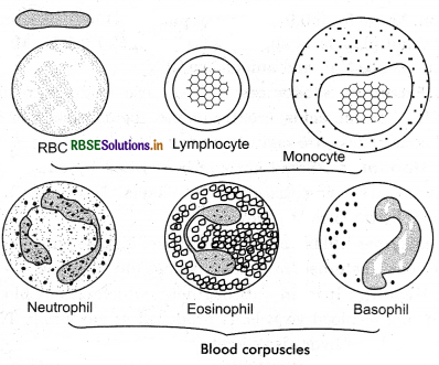
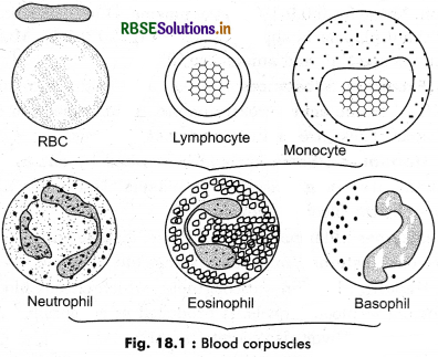
(i) Neutrophils: Their nucleus is made of 2 - 5 lobes which are connected by thin threads. These are most active WBCs and measure 10 - 12 μ in diameter. These are also known as heterophils. These are about 60 - 65% of total leucocyte count. Their granules are smaller but more numerous and can be stained by acidic or alkaline, both types of stains. Their granules are actually lysosomes and Golgy bodies. Their main function is to destroy pathogens therefore these acts as phagocytes.
(ii) Eosinophils: Their nucleus is divided into two clear lobes. Both the lobes are connected with filament. Eosinophils measure 10 - 15 μ in diameter and form 2 - 4% of total leucocyte count. These have larger granules and are stained by acidic stains like eosin. Their exact functions is not known but their number is increased many folds during allergy and infection.
(iii) Basophils: Their nucleus is made of 2 to 3 lobes or ‘S’ shaped. These are granulocytes measuring 8 - 10 μ in diameter and form 0.5 - 2% of total leucocyte count. Their granules are less numerous but relatively larger in size and can be stained with basic dyes like methylene blue. Bosophils secrete heparin and histamine. Hiparin is an anticoagulant and histamine promotes inflammatory reaction.
B. Agranulocyles: Cytoplasm of such type of WBCs is agranular and their nucleus is spherical, without lobed. These are of two types:
(i) Lymphocytes: These are smallest WBCs and measure 8 - 12 μ in diameter. These form 30% of total WBCs. Their nucleus is spherical and large in size, therefore, the cytoplasm is restricted to peripheral rim only. On the basis of their size these are grouped as small and large lymphocytes. Lymphocytes are produced in lymph glands. Their main function is to produce antibodies.
(ii) Monocytes: These are comparatively large sized and measure 12 - 15 μ in diameter. These form about 6% of total leucocyte count. These have large, horse - shoe - shaped nucleus. These are produced in lymph nodes and spleen. These are actively wondering cells engaged in phagocytosis, and are converted into macrophages after reaching tissue fluid. In tissue fluid, these dispose off as dead or worn out cells.
Question 3.
Describe blood groups found in human beings.
Answer:
There is a decrease of blood in human due to more bleeding or due to any disease then doctors demand to donate blood from any relative to patient to overcompensate blood. Before doing this they undergo matching the blood of both patient and the donor. If unmatched blood is transfused to patient, as reaching in its body, RBCs starts sticking together to form large clumps. It is called agglutination of blood. It ultimately leading to death of the patient.
Carl Landsteiner observed that the blood of all human beings is not same and the blood group of the recipient and the donor should be same for a successful transfusion. RBCs and plasma have some specific proteins, due to their interaction agglutination of blood takes place. For this discovery
Landsteiner awarded Nobel Prize.
Antigen and Antibodies of Blood: Human blood has two types of proteins:
A. Antigen or Agglutinogens: These are specific glycoproteins. These are present on membrane of RBCs. These are of two types: Antigen - A and Antigen - B.
On the basis of antigen present on RBCs, Landsteiner divided human blood into four groups:
- Blood group - A: A person having blood group A, has antigen A on RBC.
- Blood group - B: Antigen - B is present on red blood corpuscles.
- Blood group - AB: Both the antigens A and B are present on the RBCs of the person.
- Blood group - O: There is no antigen on the RBCs of a person.
B. Antibodies or Agglutinins: These are found in blood plasma. These are also of two types: anti - a and anti - b. The antigens which are not found on RBCs of human, there initially antibodies found in serum against them. For example, antigen B is not found on RBCs of human having blood group - A, hence its blood serum has antibody - a or antibody - b. Similarly antigen - A is absent on RBCs of a person having blood group B. Its serum contain antibody a or b. Both antigen A and B are present on RBCs of a person having AB blood group. No any antibody found in its blood serum. The person having blood group O lacks any type of antigen on RBCs but has both antibody - a and antibody b.
It is clear from the above description that the blood of any of the person can not transfuse to patient. For this purpose first blood group testing is required. Person of blood group A can take the blood of A and O group and can donate blood to person of A or AB blood group. Similarly, the person of blood group B can take the blood of B and O group and can donate the blood to person having B or AB blood group. The person having O blood group can not take blood of any other blood group but can donate to any other blood groups. It is called universal donor. The person having blood group AB can take the blood from any of other blood groups but can donate only to person having blood group AB. Blood group AB is called universal recipient.
Question 4.
What is Rh - factor? What is the cause of erythroblastosis foetalis? Describe.
Answer:
Rh - factor is discovered by Landsteiner and Wiener in 1940 in the blood of rabbit immunised with the blood of Rhesus monkey (Macaca rhesus). The red blood corpuscles of Rhesus monkey has an antigen which is called Rh factor. This antigen is found in the blood of about 85% human population. The people having this antigen in their blood are known as Rh+, while the people do not have this antigen are called Rh-.
There is no natural antibody of Rh antigen is found in human blood but Rh antibodies are produced in the blood plasma of a Rh- person after the exposure to Rh+ antigen. Hence, if such person receives the blood of Rh+ person second time, Rh antibody produced by earlier cause agglutination of the red blood corpuscles leading to the death of recipient.
Fisher told that three pseudoallelic genes found close to each other on a chromosome are responsible for the determination of presence or absence of Rh antigen in human blood. The dominant and recessive alleles of the genes are represented as CDE and cde respectively. Therefore, the genotype of a Rh- person would be CDE/cde. If any of the allele of these gene is present in dominant form, the person would be Rh+.
Erythroblastosis Foetalis
This is a special type of incompatibility found between a pregnant women (Rh -ve) and its foetus (Rh +ve). This is a lethal disorder and the child may die in foetal stage or just after the birth. These children are always Rh positive (Rh+). They have Rh+ father and Rh- mother. They obtain this character from their father only. During the late gestation period, the placenta becomes slightly weak and then some of the RBCs of the foetus reach into - mother’s blood circulation. Therefore, production of Rh antibodies starts in mother’s blood. As the mother is Rh-, the Rh antibodies do not affect the mother adversly. But during development when these antibodies reach into foetal blood or foetal fluid through placenta, they cause agglutination of red blood cells of foetus. This leads to death of the foetus. Generally, the first child is not seriously affected by this disorder because sufficient Rh antibodies are not produced till the birth of child. But the severity of this disorder increases in the fact uses of subsequent pregnancy.
Question 5.
What are pulse and heart rate. Give the difference between heart beat and pulse.
Answer:
Pulse: Pulse is the alternate expansion and elastic recoil of an artery with each systole. Pulse is the strongest in the arteries closet to the heart. Normal pulse rate ranges from 70 - 90 per minute.
Heart rate: Pulse per minute is called heart rate. Human heart beats 72. times per minute, this is designated as heart rate. It increases during exercise, fever, anger and fear.
Differences between heart beat and pulse:
|
Heartbeat |
Pulse |
|
1. It is the rhythmic contraction and relaxation of heart. |
1. It is the rhythmic contraction and relaxation in aorta and its main arteries. |
|
2. It is regulated by the nervous and endocrine systems. |
2. It is due to the flow of blood from the heart and is dependent on the rate of heart beat. |
|
3. One complete heart beat consists of one systole and one diastolel lasts for about 0.8 seconds. |
3. Pulse is a regular jerk of an artery. It depends on the rate of heart beat. |
Question 6.
What do you understand by open and closed circulatory system? Describe the significance of closed circulatory system.
Answer:
1. Open Blood Circulatory System: In this type of circulatory system blood is pumped by the heart into arteries. These arteries open into spaces or blood sinuses filled with blood and body fluid. Therefore, the blood circulates slowly at low pressure and freely in direct contact with cells and tissues and returns back to heart through veins. Open spaces filled with body fluid and blood actually represent body cavity calleod haemocoel. The fluid of haemocoel is called haemolymph. In such a way, is no separation between plasma of extracellular fluid and interstitial fluid in these animals.
In this system blood flow remains slow because due to having open spaces the heart can not exerts more pressure therefore no proper control or distribution of blood. Almost all arthropodes, all molluscs except cephalopods, and tunicates have open circulatory system.
2. Closed Blood Circulatory System: In this type of blood circulatory system, blood is pumped into arteries at high pressure. The arteries are divided to arterioles and arterioles divided into thin capillaries. Capillaries passing through tissues. The exchange of substances and gases between blood and interstitial fluid takes place through the walls of capillaries. These capillaries coming forward combine to form small veins and veins combine to form big veins which carry blood from tissues to heart. In such a way the blood comes in contact with cells or tissues and it remains closed therefore is called closed circulatory system. This type of circulatory system is found in many ipvertebrates like earthworm and Nereis, octopus, echinodermates etc., and all the vertebrates.
The open blood circulatory system is considered most useful because:
- It creates high blood pressure.
- It does not directly come in contact with body tissue rather comes in contact by blood capillaries.
- The distribution of blood in organs is controlled and regulated.
- The blood quickly returns into heart.
The path of closed blood circulatory, system is as follows:
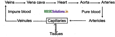
In vertebrates including mammals, these vessels of closed circulatory system reach into almost all the parts and organs of the body, but some places are also found where these vessels do not reach. Such as dermis of episkin, epithelium, cornea of eye, cartilage tissue, enamel of teeth etc. In these part, fluid with nutrients reach through interstitial fluid diffusing from blood vessels.
Circulatory System in Human
There are two types of fluids are circulated in the human body, the blood and the lymph. There are following two types of circulatory systems found in human body for circulation of blood and lymph.
- Blood Circulatory System
- Lymphatic System
1. Blood Circulatory System: All the vertebrates including human beings have closed blood vasculatory or circulatory system, which performs the function of transportation of nutrients, oxygen, hormones and all other important substances to tissues and transportation of carbon dioxide and other wastes from tissues to excretory organs.
There are following three components of the blood circulatory system:
- Pumping Organ: Heart
- Medium: Blood and Lymph
- Vessels: Arteries, Veins and Capillaries
Question 7.
What do you understand by systole and diastole?
Answer:
The heart is a muscular pump which sends the blood brought by veins to different organs of the body through the arteries by its rhythmic contraction. This regular and rhythmic contraction of heart is called heart beat. In each beat the heart contracts once and then comes in normal state. The contraction phase of the heart is called systole and relaxation phase is called diastole. The blood pumped from heart to various organs through arteries by systole while comes back from organs to heart through veins by diastole. The various events occuring in the heart from the end of one heart beat to the end of the next one, constitute cardiac cycle. In a cardiac cycle, the chambers of the heart contract and relax in a particular manner. The heart of an adult human being beats at a rate of 72 times per minute, hence, the duration of each cardiac cycle is 60/72 = 0.83 seconds.
Cardiac Cycle:
There are the following four events in the cardiac cycle:
1. Atrial Systole: During this period the blood moves to right and left ventricles through atrioventricular aperture due to the contraction of right and left atria. The duration of atrial systole is 0.1 seconds such a way both the ventricles filled with blood.
2. Ventricular Systole: During this period there is contraction in both the ventricles. Consequently the impure blood from right ventricle enters pulmonary arteries through which it reaches to lungs for oxygenation, and oxygenated (pure) blood of left ventricle is sent to different parts of the body through aorta. This phase lost for 0.3 seconds.

3. Atrial Diastole: During this period atria remain in relaxed state. In atrial diastole, the blood brought back by vena cava fill the atria. Right atrium receives blood through superior and inferior vena cava, while left atrium receives blood from pulmonary veins. Atrial diastole lasts for 0.7 seconds.
4. Ventricular Diastole: It is also called joint diastole. During ventricular relaxation, ventricles gets filled with blood received from atria. It lasts for 0.5 seconds.
A new atrial systole starts to begin a new cardiac cycle, 1/10 seconds before the completion of ventricular diastole. Out of 0.5 seconds period of ventricular diastole, for 0.4 seconds atria also remain in diastolic phase. Such a way, during a period of 0.4 seconds of a total cardiac cycle of 0.8 seconds, both atria and ventricle remain in diastolic phase. It is known as joint diastole or general pause.
Question 8.
Write the functions of the blood.
Answer:
1. Transport of Oxygen: Oxygen reaches to blood of pulmonary capillaries by diffusion. Its 2% part dissolves in plasma but mostly amount combining with haemoglobin to reaches various organs of the body. In region of higher O2 concentration. Oxygen combines with haemoglobin to form unstable compound oxyhaemoglobin.
Hb + 4O2 → Hb(O2)4
Reaching in different organs and tissues (where the concentration of O2 is less) oxyhaemoglobin breaks down into O2 and haemoglobin and O2 is received by tissues. The oxygen transportation capacity of blood is 40 times more due to presence of haemoglobin.
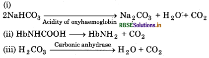
2. Elimination of CO2: CO2 produced in tissues on reaching into blood plasma combines with sodium carbonate to form sodium bicarbonate.
Na2CO3 + CO2 + H2O → 2NaHCO3
Therefore, CO2 present in blood in the form of bicarbonate and carbonic acid and carried out intolungs. On reaching into lungs various factors remove CO2:

Released CO2 diffuses into alveolar air through capillaries walls.
3. Transportation of Nutrients: Digested and absorbed food mixed up into blood plasma and carried oiit to different parts of the body.
4. Transportation of excretory substances: The substances excreted from different parts of the body are collected in blood plasma and carried out into liver and kidney. From where they are eliminated by the process of excretion.
5. Regulation of body temperature: More temperature is produced by metabolic process in different parts pf the body. Some parts of body has low temperature where they are in contact with air. Blood circulates throughly in body and regulates body temperature.
6. Transportation of other substances: Blood also transports hormones, vitamins, minerals, toxic substances, antitoxins, antibodies etc.
7. Protection from disease: Phagocytes eat pathogens. Protective cells of blood destroy pathogens producing antitoxin.
8. Clotting of Blood: At the place of injury blood forms a clot by which bleeding from wound stops. Thrombocytes of blood help in this process.
9. Healing of Wounds: For proper healing of wpunds blood carries important constituents to the injured part.
10. Creation of Pressure: Blood develops pressure in different parts of the body and maintains it.
11. Scavengiring of body: WBCs phagocytize the dead cells and remove the dead and damaged cells and waste substances.
Question 9.
Give short notes on lymph. Give the functions of lymph.
Answer:
Lymphatic system is a fluid pickup system which works with closed circulatory system. It is found in the form of an extensive network of thin walled vessels. Lymphatic system, works as an accessory drainage system for the body. At the arteriolar end of the capillaries some part of blood plasma is filtered out into the tissue fluid through capillary walls. Most of the tissue fluid re - enter the capillaries at their venular end to join the venous blood.
But as the amount of fluid which comes out through capillaries is much more than the amount of fluid which enters the venules, the excess of this outflow is supplied back to the circulatory system through lymphatic system. The fluid which enters the lymph vessels is called lymph.
The lymphatic, system of human is made of lymph, lymph vessels and lymph nodes.
1. Lymph: Lymph is a translucent, alkaline fluid found in lymph vessels and in between blood capillaries and tissues. It is a colourless liquid resembling plasma. But it, has comparatively very less amount of proteins, calcium and phosphorus. It has much amount of excretory materials. Lymph lacks thrombocytes and RBCs. It has lymphocytes and leucocytes in more numerous.
2. Lymph Vessels: Lymph capillaries unite to form lymph vessels which are like as veins in their structure. These are thin walled having semilunar valves like veins. There is no any pumping station found in lymphatic system but the lymph flows in lymph vessel by the action of circular and longitudinal involuntary muscles present in the walls of the lymph vessels.
The lymph vessels of left side of neck and head, left forearm, both the legs, thorax and different organs of abdominal cavity opens into a big left thoracic lymph duct. This vessel is attached to a sac-like structure which is called cisterna chyli, lies below the diaphragm in the abdominal cavity. It goes upwards and pierces the diaphragm through aortic hiatus and drain into left subclavian vein.
Similarly, the lymph vessels of right forearm and head, neck and right side of thorax opens into a right thoracic duct. It is smaller than that of left thoracic lymphatic duct and drain into right subciavian vein. The lymph capillaries of villi found on inner lining of small intestine are called lacteals. These lacteals absorb fats from small intestine.
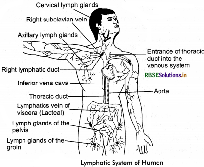
3. Lymph Nodes: Lymph nodes represent aggregates of lymphoid tissue present in the route of lymphatic vessels. The lymph of the lymph vessels passes through these lymph nodes. These lymph nodes are mainly located in neck, arm pit, thorax, abdomen, groin and near large blood vessels. Tonsils are also lymph nodes.
Functions of Lymphatic System
The lymphatic system:
- filters bacteria, foreign materials, toxins and any harmful material.
- drains away excess fluid to prevent water clogging of the tissues and cells.
- transports proteins back into the blood supply.
- produces lymphocytes which protects and defend the body against infection.
- produces antibodies to fight bacteria.
- absorbs fat from the intestine and transport it to the liver.
Question 10.
How many types of heart on the basis of origin of heart beat? Explain.
Answer:
On the basis of origin of heart beat the hearts can be grouped in the following two types:
1. Neurogenic heart: In this type of heart, the heart beat originates through nervous tissue. Most of the invertebrate animals have neurogenic heart.
2. Myogenic heart: Heart beat originates through a special muscular tissue in this type of heart. This tissue does not have contractibility but it has the ability to generate motor impulse by self excitation, and transmit it as a cardiac impulse. In this type of heart, neural or hormonal stimuli do not generate cardiac impulse (heart beat) but affect the rate of heart beat. Therefore, heart beat continues even if the nervous connection of heart is removed. This type of heart is found in molluscs and vertebrates.
Question 11.
What are tonsils? Give their types.
Answer:
Tonsils: Tonsils are lobes of lymphoid tissues which are enclosed in a capsule of connective tissue. There are three types of tonsils:
- Pharyngeal tonsils: They are situated in the upper posterior wall of pharynx. Pharyngeal tonsils are also known as Luschka’s tonsils.
- Palatine tonsils: They are present between the columns of fauces on both sides of pharynx.
- Lingual tonsils: Lingual tonsils are found at the root of the tongue.
- Tonsils help to fight infections.
Question 12.
Write the differences between sino - atrial node and atrio - ventricular node.
Answer:
Differences between sino - atrial node and atrio - ventricular node.
|
Sino - atrial node (SA node) |
Atrio - ventriculai node (AV node) |
|
1. Sino - atrial node is located in superior lateral wall pening of superior vena cava. |
1. AV - node is present in the posterior septal wall of right atrium near the opening of coronary sinus. |
|
2. It is longer. |
2. It is shorter. |
|
3. It generate the cardiac impulse. |
3. It relays and intensifies the cardiac impulse. |
|
4. SA node transmits impulse directly to the two atria. |
4. AV node carries impulse to the two ventricles through AV bundle, its branches end terminal strands. |
|
5. SA node acts as pace maker. |
5. It is influenced by SA node. |
|
6. Autorhythmic fibres initiate cardiac action potential, which set basic pace for heart rate and conduct throughout both atria. |
6. Receives action potentials from SA node and posses them to AV bundle. |
Long Answer Type Questions
Question 1.
Write about the components of plasma.
Answer:
Plasma is pale coloured, slightly alkaline clear, transparent, non - living fluid. It constitute about 55 - 60% of the blood. Plasma contains following components:
Water: 90 - 92%
Inorganic substances: 1%
Plasma proteins: 6 - 8%
Other organic substances: 1%
Chlorides, carbonates, bicarbonates, sulphates and phosphates of sodium, potassium, calcium and magnesium are main inorganic substances found in plasma. Among organic substances plasma proteins like albumins, globulins, fibrinogens and prothrombin constitute about 6 - 8% part of plasma. All these proteins are soluble and present in colloidal form in plasma. Therefore, these make plasma viscous and regulate osmotic pressure. Prothrombin and fibrinogen proteins helps in coagulation of blood and forms 4% of plasma proteins. Albumin forms 50% of all plasma proteins and these maintain osmotic pressure of the blood. Globulins form 43% of total plasma proteins.
These are of three types: α - globulins, ß - globulins and γ - globulins. α and ß globulins transport proteins to different parts of the body, while γ - globulins protect the body from infections. Plasma also contains ions such as Na+, Ca++, Mg++, HCO3- , Cl- etc. Plasma also contain glucose, amino acids and lipids due to any infection in the body. The blood clotting factors also present in plasma in inactive state. Plasma without clotting factor is called serum.
1. Plasma Proteins: These constitute about 70% part of plasma. These are of following type:
- Globulins or Immunoglobulins: These proteins form about 43% part of total plasma proteins. These are of three types: Alpha (α), Beta (ß) Gamma (γ) globulins. Alpha and beta globulins transport proteins to different parts of the body while gamma globulins protects the body from infections.
- Albumins: These proteins form about 53% of all plasma proteins. These maintain osmotic pressure of the blood.
- Fibrinogen and prothrombin: These form about 4% of plasma proteins. These help in coagulation of blood.
2. Organic and Digested Materials: Plasma contains carbohydrates (glucose, fructose, lactose), fats, fatty acids, amino acids, glycerol, vitamins, cholesterol, phospholipids, etc., in digested or undigested form. Plasma contain 80 - 100 mg glucose/100 mL blood and 0. 2% fat. Cholesterol is present about 50 - 180 mg/100 mL blood.
3. Salts: These are chlorides, carbonates, bicarbonates, sulphates and phosphates of sodium, potassium and iron. These form 0.9 - 1% part of plasma. Due to presence of them plasma is slightly alkaline (pH 7.3 - 7.5). Main cations are Na+ and anions are Cl-.
4. Excretory Substances: Urea, uric acid, ammonia (NH3), amino acids, creatinine etc., being dissolved in plasma. These are main excretory substances.
5. Hormones: Some hormones secreted by endocrine glands are being dissolved in plasma. Hormones are carry by the blood to their targets.
6. Enzymes: Plasma contains lipase, distase, glucolase, protease, esterase, nuclease, oxidase etc., enzyme.
7. Heparin: It is an anticoagulent, which checks blood clotting in blood vessels. It produced in basophils. The mast cells of liver also secrete heparin.
8. Antigen and Antibodies: Bacteria and external proteins reaching in blood are called antigens. Plasma produces special type of proteins to reduce the effect of these antigens. These proteins are called antibodies. They are inactive antigens.
9. Gases: The gases such as carbon dioxide, oxygen, nitrogen etc., are also dissolved in plasma.
10. Drugs: The drugs taken by the person also present in plasma.
Functions of Plasma:
- Plasma transports various nutrients, hormones, enzymes and excretory substances from one place to another.
- Maintains fluid quantity of blood.
- Heparin present in plasma do not allow to coagulate blood in vessels.
- Its fibrinogen protein forms blood clot after injury.
- The globulin protein acts as immunity.
- It maintains pH of blood.
- It regulates body temperature.
- It removes excretory substances along with CO2 from body.
Question 2.
Write short notes on the following:
(i) Erythrocytes
(ii) Leucocytes
(iii) Platelets.
Answer:
(i) Red Blood Corpuscles (RBCs) or Erythrocytes: These are most in number than other blood cells. There are about 5 million/mm3 of RBCs in the blood of an adult healthy human male. In human females their nurtiber is about 4.5 million/mm3. During foetal stage RBCs are produced in the liver and spleen but after the birjfet this function is taken over by red bone marrow of long bones.
RBCs are oval, biconcave and nucleated in vertebrates from fishes to birds and in mammals like camel and llama. In all other mammals, the RBCs are circular, biconcave and enucleated. They are nucleated during their initial stages of development but later on along with nucleus other cell organelles like mitochondria, endoplasmic reticulum. Golgi apparatus and ribosomes etc., also disappear. Biconcave shape increases the surface area while absence of organelles provide extra space to accomodate haemoglobin.
Human RBCs measure 7.2 - 7.5 µ in diameter and 2.2 µ in thickness and have an average volume of 88 - 90 µ3. Process of their production is termed as erythropoiesis. The maturation of RBCs is controlled by folic acid and vitamin B12, therefore lack of these vitamins causes anaemia. Red blood cells presence in excess are stored in spleen, hence it is called blood bank of the body. Apart from this spleen is called grave yard of RBCs as it removes dead cells or worn out cells. RBCs have a life span of 120 days. The dead and worn out RBCs are destroyed by reticuloendothelial cells of bone marrow, liver and spleen.
(ii) White Blood Corpuscles (WBCs) or Leucocytes:
These are circular, amoeboid or irregular shaped, colourless, nucleated wondering cells. These have the capacity to squeez out from blood capillaries into tissue fluid where these destroy pathogens and foreign antigens to protect the body. There are about 6000 - 8000 WBCs/mm3 blood in a healthy adult man. They are produced in bone marrow, Peyer’s patches of intestinal mucosa, lymph nodes, thymus and spleen. The production of WBCs is called leucopoiesis. The average life span of WBCs is 4 - 7 days.
There are the following two groups of white blood corpuscles:
- Granulocytes
- Agranulocytes .
(iii) Blood Platelets: These blood corpuscles are also called thrombocytes. These are found in the blood of mammals only. Their number is 2 - 4 lac per mm3. Platelets are oval or circular plasma elements measuring 2 - 5 µ in diameter. They are formed by the fragmentation of megakaryocytes of bone marrow. These do not have nucleus. This feature differentiates them from thrombocytes of lower vertebrates. Platelets have a life span of 2 - 5 days. Many platelet factors and enzyme thromboplastin are released by damaged or injured platelets, which help in blood clotting. Apart from this platelets also secrete a substance serotonin which inhibits the haemorrhagic effects by vasoconstriction of blood vessels.
Question 3.
Write the functions of blood.
Answer:
1. Transport of Oxygen: Oxygen reaches to blood of pulmonary capillaries by diffusion. Its 2% part dissolves in plasma but mostly amount combining with haemoglobin to reaches various organs of the body. In region of higher O2 concentration. Oxygen combines with haemoglobin to form unstable compound oxyhaemoglobin.
Hb + 4O2 → Hb(O2)4
Reaching in different organs and tissues (where the concentration of O2 is less) oxyhaemoglobin breaks down into O2 and haemoglobin and O2 is received by tissues. The oxygen transportation capacity of blood is 40 times more due to presence of haemoglobin.
2. Elimination of CO2: CO2 produced in tissues on reaching into blood plasma combines with sodium carbonate to form sodium bicarbonate.
Na2CO3 + CO2 + H2O → 2NaHCO3
Therefore, CO2 present in blood in the form of bicarbonate and carbonic acid and carried out intolungs. On reaching into lungs various factors remove CO2:
Released CO2 diffuses into alveolar air through capillaries walls.
3. Transportation of Nutrients: Digested and absorbed food mixed up into blood plasma and carried oiit to different parts of the body.
4. Transportation of excretory substances: The substances excreted from different parts of the body are collected in blood plasma and carried out into liver and kidney. From where they are eliminated by the process of excretion.
5. Regulation of body temperature: More temperature is produced by metabolic process in different parts pf the body. Some parts of body has low temperature where they are in contact with air. Blood circulates throughly in body and regulates body temperature.
6. Transportation of other substances: Blood also transports hormones, vitamins, minerals, toxic substances, antitoxins, antibodies etc.
7. Protection from disease: Phagocytes eat pathogens. Protective cells of blood destroy pathogens producing antitoxin.
8. Clotting of Blood: At the place of injury blood forms a clot by which bleeding from wound stops. Thrombocytes of blood help in this process.
9. Healing of Wounds: For proper healing of wpunds blood carries important constituents to the injured part.
10. Creation of Pressure: Blood develops pressure in different parts of the body and maintains it.
11. Scavengiring of body: WBCs phagocytize the dead cells and remove the dead and damaged cells and waste substances.
Question 4.
Mention the features of human circulatory system. Write functions of circulatory system.
Answer:
Features of human circulatory system:
- Two types of fluids are circulated in the body of human beings, the blood and the lymph. For their circulation there are following two types of circulatory systems: blood circulatory system and lymphatic system.
- The blood circulatory system is made of blood, heart and vessels. Vessels are further divided into arteries, arterioles, veins, venules and capillaries.
- The lymphatic system is made of veins, venules, capillaries, lymph and lymph nodes.
- Human circulatory system is closed type where, blood flows in enclosed vessels.
- In closed circulatory system tissues and cells never come in contact with blood.
- In blood circulatory system, the heart acts as pumping station.
- In circulatory system blood flows quite rapid while lymph flows quite slow.
Functions of Circulatory system:
- Transportation of nutrients, oxygen, carbon dioxide, waste products, hormones and other materials.
- It regulates body temperature.
- It maintains homeostasis.
- It helps in protection of body against disease.
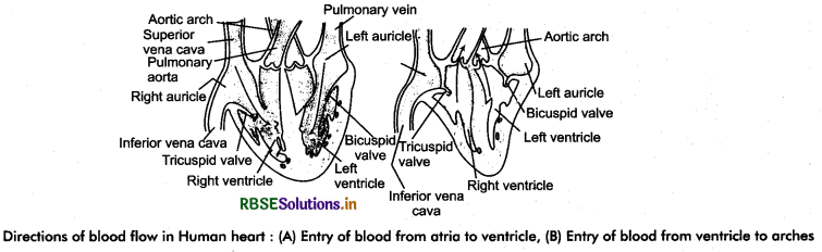
Question 5.
Explain the external and internal structure of heart.
Answer:
Heart is a busiest organ functioning as pumping station. It pumps blood into whole body parts. It is situated in between both lungs in mediastinum part of thorax, on ventral side, just above the diaphragm and slightly tilted towards left. Therefore, its one third part is present towards the right side of mid line of the body, while two third part is present towards left sides. The broader part of the heart located towards right side is called its base. It tilted towards back or dorsal side. The lower pointed end of the heart forms its apex, which is tilted towards left and front or ventral side.
External Structure of Human Heart:
The heart of human being is a muscular, conical, hollow and pulsatile organ. It is pinkish in colour and is about the size of our fist. It measures 12 cm in length and 9 cm in width. Its enteroposterior diameter is about 6 cm. Its weight varies in males from 280 - 340 g and in females from 230 - 280 g. The heart is enclosed in a fibrous sac known as the pericardium. The pericardium is made up of two layers: The outer fibrous pericardium and the inner serous pericardium. The outer pericardium is made of fibrous tissues and at the upper side it remains in continuity with the tunica adventia of larger blood vessels of the heart.
On lower side, it is closely applied to diaphragm. Serous pericardium consists of two layers outer perietal membrane which lines fibrous pericardium and inner epicardium which is closely applied on heart muscles to form its outer covering. The space between parietal membrane and epicardium is called pericardial cavity which filled with pericardial fluid. This fluid prevents friction during beating of heart and protects the heart from mechanical shocks.
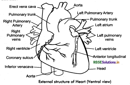
Human heart is four chambered, consisting of two atria and two ventricles. The left and right atria are separated externally by a shallow vertical interatrial groove. The atria are demorated externally from the ventricles by an oblique groove called atrioventricular sulcus. There are also present coronary sulcus, anterior interventricular sulcus and posterior interventricular sulcus. These have coronary arteries, through which the heart receives blood.
The left atrium is smaller than the right atrium. Each atrium has an appendages called an auricle which increases its surface area. The superior vena cava, inferior vena cava and coronary sinus opens into right atrium. The left atrium receives four openings of pulmonary veins. Ventricles are thick walled. The left ventricle is longer and narrower than the right ventricle. Its walls are about three times thicker than the right ventricle. The pulmonary trunk arises from the right ventricle. The aorta arises from the left ventricle.
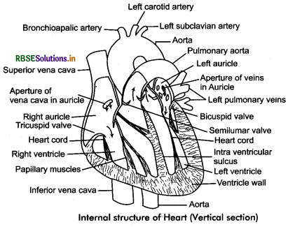
Internal structure of the Heart:
Structure of the heart wall: There are following three layers found in the wall of the, heart:
1. Epicardium: It is the outermost layer of the heart which made of visceral serous membrane of the serous pericardium. It rests on a basment membrane and represents mesothelium.
2. Myocardium: It is the middle thick layer of the heart, made of cardiac muscles cardiac muscle fibres are striated and branched but involuntary in nature. Myocardium is thicker at the apex of the heart and gradually becomes thin towards its base.
3. Endocardium: It lines the cavity of the heart and made of flat squamous cell derived from endoderm of the embryo. This endothelium rests on a basement membrane.
The cavity of the heart is divided into four chambers internally also. Right and left atria are present on the upper side while right and left ventricles form the lower side of the heart.
Atrium: There are two atria in the heart which are separated to each other by an interatrial septum into right atrium and left atrium. During foetal stage there is an aperture in the interatrial septum called faramen ovalis. It closes automatically after the birth, and is, represented as a depression called fossa ovalis, on the right side of interatrial septum.
The right atrium receives blood from superior vena cava, inferior vena cava and coronary sinus. For this purpose right atrium has openings. Through these apertures impure blood from the upper and lower sides of the body reaches right atrium.
For pulmonary veins, bringing oxygenated blood from the lungs opens into left atrium separately through four openings. Left and right atria collect the blood and pump it to left and right ventricles respectively. As atria simply collect the blood, they are thin walled.
Ventricles: Ventricles are divided into left and right ventricle by an interventricular septum. Right ventricle contracts to pump the deoxygenated blood into lungs while left ventricle contracts to pump oxygenated blood to different parts of the body. Therefore, the walls of the
ventricles are comparatively thicker than the walls of the atria. The wall of the ventricles is elevated in the form of small irregular folds called trabeculae carnae. Apart from this inner sides of ventricles also have muscular folds called papillary muscles or columnae carnae.
Question 6.
What is cardiac cycle? Write its various components.
Answer:
Cardiac Cycle:
There are the following four events in the cardiac cycle:
1. Atrial Systole: During this period the blood moves to right and left ventricles through atrioventricular aperture due to the contraction of right and left atria. The duration of atrial systole is 0.1 seconds such a way both the ventricles filled with blood.
2. Ventricular Systole: During this period there is contraction in both the ventricles. Consequently the impure blood from right ventricle enters pulmonary arteries through which it reaches to lungs for oxygenation, and oxygenated (pure) blood of left ventricle is sent to different parts of the body through aorta. This phase lost for 0.3 seconds.

Question 7.
Write the working mechanism of the human heart.
Answer:
The heart is a muscular pump which sends the blood brought by veins to different organs of the body through the arteries by its rhythmic contraction. This regular and rhythmic contraction of heart is called heart beat. In each beat the heart contracts once and then comes in normal state. The contraction phase of the heart is called systole and relaxation phase is called diastole. The blood pumped from heart to various organs through arteries by systole while comes back from organs to heart through veins by diastole. The various events occuring in the heart from the end of one heart beat to the end of the next one, constitute cardiac cycle. In a cardiac cycle, the chambers of the heart contract and relax in a particular manner. The heart of an adult human being beats at a rate of 72 times per minute, hence, the duration of each cardiac cycle is 60/72 = 0.83 seconds.
Cardiac Cycle:
There are the following four events in the cardiac cycle:
1. Atrial Systole: During this period the blood moves to right and left ventricles through atrioventricular aperture due to the contraction of right and left atria. The duration of atrial systole is 0.1 seconds such a way both the ventricles filled with blood.
2. Ventricular Systole: During this period there is contraction in both the ventricles. Consequently the impure blood from right ventricle enters pulmonary arteries through which it reaches to lungs for oxygenation, and oxygenated (pure) blood of left ventricle is sent to different parts of the body through aorta. This phase lost for 0.3 seconds.
3. Atrial Diastole: During this period atria remain in relaxed state. In atrial diastole, the blood brought back by vena cava fill the atria. Right atrium receives blood through superior and inferior vena cava, while left atrium receives blood from pulmonary veins. Atrial diastole lasts for 0.7 seconds.
4. Ventricular Diastole: It is also called joint diastole. During ventricular relaxation, ventricles gets filled with blood received from atria. It lasts for 0.5 seconds.
A new atrial systole starts to begin a new cardiac cycle, 1/10 seconds before the completion of ventricular diastole. Out of 0.5 seconds period of ventricular diastole, for 0.4 seconds atria also remain in diastolic phase. Such a way, during a period of 0.4 seconds of a total cardiac cycle of 0.8 seconds, both atria and ventricle remain in diastolic phase. It is known as joint diastole or general pause.
Question 8.
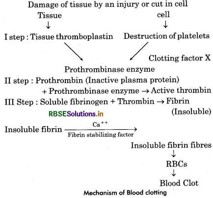
Write the mechanism of blood clotting.
Answer:
Blood leaking out from the blood vessels on the injured part or wound or from a cut in the body gets clotted within 3 to 6 minutes and bleeding is stopped by formation of a jelly like substance. It is known as blood clotting or blood coagulation. Howell first described this process. After clotting a yellow coloured fluid called serum ooze out from place of injury. Serum lacks fibrinogen therefore it does not coagulate.
Mechanism of Blood Clotting:
This is a complex process which was first studied in detail by Howell. In blood an inactive protein enzyme prothrombin is found in blood. This is controlled by anticoagulant or antiprothrombin. Due to presence of heparin blood remain in fluid state and in normal state circulates in the vessels. Followings are main process occuring - in clotting of blood:
(i) Formation of Prothromboplastin or Prothrom - binase Enzyme: After getting an injury tissues release prothromboplastin a lipoprotein. The blood platelets of the same region form platelet factor III. This substance combine with calcium ions and few proteins make pro - thromboplastin and prothrombinase enzyme.
(ii) Formation of thrombin: Prothromboplastin is converted into thromboplastin with the help of Ca++. Thromboplastin reacts with inactive prothrombin and converts it into active thrombin, this process is carried out in the presence of Ca++ ions.
(iii) Formation of Fibrin fibres: Active thrombinase converts insoluble fibrin into soluble fibrinogen protein. Fine long solid fibres of fibrin form a network around the wound. In formation of this bond many RBCs and WBCs get entrapped into the network and form a clot. After some time clot starts to shrink and a light yellow coloured serum comes out. In fact serum has plasma without corpuscles. It has blood protein and minerals but lacks fibrinogen. After drying of it blood forms a complete clot and bleeding is checked completely. In some persons bleeding does not stop due to lack of clotting of blood. Therefore, person dies due to more bleeding. It is a disease called haemophilia. It is a hereditary disorder.
The following 13 coagulation factors takes part in the process of blood coagulation or blood clotting:
- Fibrinogen.
- Tissue prothrombin
- Thromboplastin
- Calcium Ions (Ca++)
- Labile factor, proaccelerin
- Hypothetical Factor, Accelerin.
- Stable Factor, Proconvertin
- Antihaemophilic Factor, AHF
- Christmas factor or plasma thromboplastin component, PTC.
- Stuart: Prower Factor.
- Plasma thromboplastin anticedent, PTA.
- Hageman Factor.
- Fibrin Stabilizing Factor or Laki Lorand Factor. Out of these factors I, II, V, VII, VIII, IX and X are synthesized in the liver.
Question 9.
What do you understand by blood groups? Explain.
Answer:
There is a decrease of blood in human due to more bleeding or due to any disease then doctors demand to donate blood from any relative to patient to overcompensate blood. Before doing this they undergo matching the blood of both patient and the donor. If unmatched blood is transfused to patient, as reaching in its body, RBCs starts sticking together to form large clumps. It is called agglutination of blood. It ultimately leading to death of the patient.
Carl Landsteiner observed that the blood of all human beings is not same and the blood group of the recipient and the donor should be same for a successful transfusion. RBCs and plasma have some specific proteins, due to their interaction agglutination of blood takes place. For this discovery
Landsteiner awarded Nobel Prize.
Antigen and Antibodies of Blood: Human blood has two types of proteins:
A. Antigen or Agglutinogens: These are specific glycoproteins. These are present on membrane of RBCs. These are of two types: Antigen - A and Antigen - B.
On the basis of antigen present on RBCs, Landsteiner divided human blood into four groups:
- Blood group - A: A person having blood group A, has antigen A on RBC.
- Blood group - B: Antigen - B is present on red blood corpuscles.
- Blood group - AB: Both the antigens A and B are present on the RBCs of the person.
- Blood group - O: There is no antigen on the RBCs of a person.
B. Antibodies or Agglutinins: These are found in blood plasma. These are also of two types: anti - a and anti - b. The antigens which are not found on RBCs of human, there initially antibodies found in serum against them. For example, antigen B is not found on RBCs of human having blood group - A, hence its blood serum has antibody - a or antibody - b. Similarly antigen - A is absent on RBCs of a person having blood group B. Its serum contain antibody a or b. Both antigen A and B are present on RBCs of a person having AB blood group. No any antibody found in its blood serum. The person having blood group O lacks any type of antigen on RBCs but has both antibody - a and antibody b.
It is clear from the above description that the blood of any of the person can not transfuse to patient. For this purpose first blood group testing is required. Person of blood group A can take the blood of A and O group and can donate blood to person of A or AB blood group. Similarly, the person of blood group B can take the blood of B and O group and can donate the blood to person having B or AB blood group. The person having O blood group can not take blood of any other blood group but can donate to any other blood groups. It is called universal donor. The person having blood group AB can take the blood from any of other blood groups but can donate only to person having blood group AB. Blood group AB is called universal recipient.
Question 10.
Mention the internal structure of artery. Write difference between arteries and veins.
Answer:
Human has closed blood circulatory system. It means that the blood is restricted to blood vessels only. To perform this function there are two systems of blood vessels found in the body - arterial system and venous system. Arteries carry blood from heart to different organs and tissues. Veins bring the blood back from organs and tissues to the heart. The blood pumped by the heart enters the aorta. Aorta divided into arteries, arterioles and fine capillaries respectively. From capillaries blood goes to venules and ultimately reaches to heart through veins and vena cava.
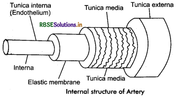
Histology of Vessels: There are three layers found in walls of arteries and veins:
- Tunica externa
- Tunica media
- Tunica interna.
1. Tunica externa: It is outermost layer of loose connective tissue. It has longitudinally arranged collagen fibres, elastic fibres and few fibroblasts. Tunica externa is found merged with the surrounding connective tissue. It has nerve also. It is more thick in arteries than that of veins and absent in capillaries.
2. Tunica media: This is the middle layer of smooth or unstriated muscles. It is covered by an elastic membrane. Muscle cells are arranged circumferentially. Tunica media is thickest layer and the thickness of layers of muscles fibres depends on the diameter of the artery. It is thicker in arteries and thinner in
veins.
3. Tunica Interna: All the structural units of this layer are arranged along its length. This layer itself consists of two layers. The inner most layer is made of endothelial cells. Just beneath this, there is a thin membranous layer made of elastic tissue. Its innermost surface is highly smooth. The capillaries have only tunica interna. The tunica media of aorta and large arteries have more elastic tissue than smaller arteries and arterioles, which have comparatively more muscular tissue.
Arteries: Arteries carry the blood pumped by heart to different organs and tissues. Arteries are thick walled vessels and branched continuously starting from aorta and pulmonary artery to the capillaries. As the move away from the heart these divide and their caliber reduces gradually. Through the diameter of the successive branches reduces gradually, the sum of the transverse section of all the branches increases. In this way the flow of blood at capillary level becomes very slow. It provides enough time for the exchange of materials with the tissues. Due to having thick walls and narrow lumen, arteries do not collapse when there is no blood in them.
Generally, arteries feel pinkish in colour and situated deep ip body than that of veins. But in wrist, neck etc., these are situated near the skin. At these places pulsation is observed in them.
Arterioles: As reaching different organs arteries divide continuously (6 to 8 time) to form thin arteries which are called arterioles. Their wall is made of mainly unstriated muscles. The squamous cells of endothelial layer of them are more flat. Due to having vasodilation and vasoconstriction capacity arteries play important role in increase or decrease blood supply as requirement. Therefore, arterioles are called stop cocks of circulation.
Capillaries: After reaching into tissues the arterioles divide into many fine branches to from capillaries. Their walls lack tunica externa and tunica media. Capillaries are of equal diameter and form a network. The endothelium of some capillaries has very fine apertures or fenestra. Different materials are exchanged between blood and tissue fluid through capillaries by simple diffusion.
Veins and Venules: The capillaries of the veins unite together to form venules in the tissue. Venules unite to form veins. As they move towards heart they form larger veins which unite to form vena cava. Superior vena cava collects blood from upper parts of the body while inferior vena cava collects blood from lower regions of the body.
Like arteries, the wall of veins is also made of three layers as described earlier, but the borders of these layers are not clear. Due to lesser muscular and elastic tissue in the tunica media, the walls of the veins are comparatively thinner and collapsible. They have lesser toughness and elasticity also. Therefore, during a cut or any other injury the veins collapse.
As the walls of the veins are comparatively thinner, these have wider lumen. The speed of blood flow, is very slow and blood pressure is also low in vein. Therefore, the veins of distal or peripheral parts of the body have semilunar valves, which allow the blood to flow in one direction only. The network of fine blood vessels supplying blood to the blood vessels is called vasa - vasorum.
|
Arteries |
Veins |
|
1.These moves away from the heart. |
1. These moves towards the heart. |
|
2. These distribute blood to the body organs. |
2. These collect blood from body organs. |
|
3. Blood pressure is high in arteries. |
3. Blood pressure in veins is low. |
|
4. Valves are absent. |
4. Valves are present. |
|
5. These carry oxygenated blood except pulmonary artery. |
5. These carry deoxygenated blood, except pulmonary vein. |
|
6. Arteries end in capillaries. |
6. Veins starts in blood capillaries. |
|
7. These are deep seated. |
7. These can be seen subcut - eneously. |
|
8. Arteries are further divide into arterioles. |
8. Veins are further divide into venules. |
|
9. These are round and relatively thick walled. |
9. Veins |
|
10. Arteries have narrow lumen. |
10. Veins have large lumen. |
|
11. They do not collapse when there is no blood in them. |
11. These collapse when there is no blood in them or cut across. |
|
12. Arteries reddish or pinkish in colour. |
12. Veins are bluish in colour. |
|
13. Arteries show spurty movement of blood giving pulse. |
13. Veins show sluggish movement of blood. |
|
14. If arterial wall is injured, the blood comes out like fountain in a large area all around the artery. |
14. If venous wall is injured, blood comes out, collects in a pool in a small area around vein. |
Question 11.
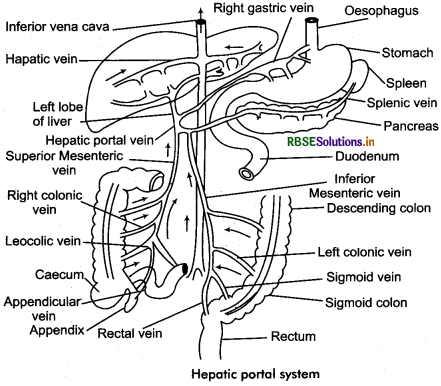
Explain hepatic portal system.
Answer:
Most of the veins carry the venous blood collected from capillaries directly to the heart, but in some cases the veins bringing venous blood enter a specific organ and form a network of small veins and united with the network of capillaries of that organ. Such veins are called portal veins and their system is known as portal system. The hepatic portal system is an example of portal system. Other example of portal system is renal portal system.
All mammals including human have only hepatic portal system which starts from alimentary canal and ends in liver. It has following veins:
- Splenic vein: Collect blood from spleen.
- Superior mesenteric vein: It collects blood from small intestine, caecum and descending colon.
- Oastric vein: It collects blood from stomach.
- Pancreatic vein: It collects blood from pancreas.
- Duodenal vein: It collects blood from duodenum.
- Cystic vein: It collects blood from gall bladder.
- Inferior mesenteric vein: It collects blood from colon, sigmoid colon and rectum.
These all veins unite to form a hepatic portal vein which opens into left lobe of liver. On reaching into liver it divides into capillaries. These capillaries now unite to form veins which open into inferior vena cava.
Importance of Hepatic Portal System:
- It regulates the amount of sugar in body.
- It destroys bacteria and pathogens come with food.
- It inactivates toxic substances.
- It converts ammonia obtained from deamination of amino acid into urea.
- It induces glyconeogenesis on deficiency of sugar in blood.
Question 12.
Describe double circulatory system.
Answer:
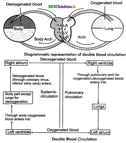
Double Blood Circulation:
In human there are two circuits of blood circulation for greater efficiency and completely prevent the mixing of oxygenated and deoxygenated blood. Usually it is called double blood circulation. Double circulation is the passage of same blood twice in the heart through separate pathways for completing one cycle, it consists of pulmonary circulation and systemic circulation.
Pulmonary circulation is the movement of blood from heart to the lungs, and back to the heart again. Deoxygenated blood is pumped out of the right ventricle of the heart into the pulmonary trunk, then to the pulmonary arteries and into the lungs via pulmonary veins. Oxygenated blood is then drained into the left atrium. Blood is then circulated in the systemic circulation. The flow of oxygenated blood from the left ventricle to all parts of the body and deoxygenated blood from various body parts to the right atrium is called systemic circulation.
Significance of Double Blood Circulation:
The advantage of double blood circulation is that the blood can be sent to the lungs to pick up oxygen and then returned to the heart to be pumped again before travelling around the body. The blood therefore is pumped through the capillary bed (which slows down and reduces its pressure) then receives another pump before it enters another capillary bed. Double Circulatory systems are therefore high pressure system. In this type of circulation there is no mixing of the oxygen rich blood and oxygen poor blood in the heart. Oxygenated blood carries more oxygen to different body parts as well as more CO2 is carried with deoxygenated blood for the removal through lungs.

- RBSE Solutions for Class 11 Biology Chapter 10 Cell Cycle and Cell Division
- RBSE Solutions for Class 11 Biology Chapter 9 Biomolecules
- RBSE Solutions for Class 11 Biology Chapter 8 Cell: The Unit of Life
- RBSE Solutions for Class 11 Biology Chapter 7 Structural Organisation in Animals
- RBSE Solutions for Class 11 Biology Chapter 6 Anatomy of Flowering Plants
- RBSE Solutions for Class 11 Biology Chapter 5 Morphology of Flowering Plants
- RBSE Solutions for Class 11 Biology Chapter 4 Animal Kingdom
- RBSE Solutions for Class 11 Biology Chapter 3 Plant Kingdom
- RBSE Solutions for Class 11 Biology Chapter 2 Biological Classification
- RBSE Solutions for Class 11 Biology Chapter 1 The Living World
- RBSE Solutions for Class 11 Biology Chapter 5 पुष्पी पादपों की आकारिकी