RBSE Class 11 Biology Important Questions Chapter 17 Breathing and Exchange of Gases
Rajasthan Board RBSE Class 11 Biology Important Questions Chapter 17 Breathing and Exchange of Gases Important Questions and Answers.
Rajasthan Board RBSE Solutions for Class 11 Biology in Hindi Medium & English Medium are part of RBSE Solutions for Class 11. Students can also read RBSE Class 11 Biology Important Questions for exam preparation. Students can also go through RBSE Class 11 Biology Notes to understand and remember the concepts easily.
RBSE Class 11 Biology Chapter 17 Important Questions Breathing and Exchange of Gases
Multiple Choice Questions
Question 1.
The tidal volume in resting stage in a normal human is:
(a) 0.5 liter
(b) 2.5 liter
(c) 1.2 liter
(d) 4.9 liter
Answer:
(a) 0.5 liter

Question 2.
What is responsible for transport of O2 from alveolus to blood in capillaries of lungs?
(a) Active transport
(b) Filteration
(c) Facilitated diffusion
(d) Passive diffusion
Answer:
(d) Passive diffusion
Question 3.
Which structure is final dividation of lung and is site of gaseous exchange?
(a) Trachiole
(b) Air alveolus
(c) Trachea
(d) Bronchioles
Answer:
(b) Air alveolus
Question 4.
Carbon dioxide is transported through:
(a) Haemoglobin
(b) Plasma
(c) RBCs
(d) All of these
Answer:
(d) All of these
Question 5.
Breathing rate is increased in running , because blood contains:
(a) less amount of lactic; acid
(b) more concentration of CO2
(c) less concentration of CO2
(d) less concentration of CO2 and lactic acid
Answer:
(b) more concentration of CO2
Question 6.
Enzyme used for the transport of CO2 in blood:
(a) carboxylase
(b) carboxykinase
(c) carbonic anhydrase
(d) none of these
Answer:
(c) carbonic anhydrase
Question 7.
The respiratory centre present in:
(a) cerebellum
(b) ceribrum
(c) medula oblongata
(d) hypothalamus
Answer:
(c) medula oblongata
Question 8.
Book lungs are the respiratory organ of:
(a) protozoans
(b) cnidarians
(c) arthropods
(d) amphibians
Answer:
(c) arthropods
Question 9.
Gills are the respiratory organs of:
(a) Birds
(b) Fishes
(c) Insects
(d) Mammals
Answer:
(b) Fishes
Question 10.
Percentage of carbon dioxide transported by red blood cells is:
(a) 70%
(b) 20 - 25%
(c) 97%
(d) 7%
Answer:
(b) 20 - 25%
Question 11.
About 70% of CO2 is transported by blood to lungs in the form of:
(a) Carbaminohaemoglobin
(b) Bicarbonate ions
(c) Dissolved gaseous molecules
(d) Linkage with RBC
Answer:
(b) Bicarbonate ions
Question 12.
When temperature is low, then oxyhaemoglobin curve will be:
(a) more bend
(b) upright
(c) parabolic
(d) all of these
Answer:
(a) more bend
Question 13.
The enzyme carbonic anhydrase takes part in CO2 transport, found in:
(a) WBCs
(b) Lymphocytes
(c) RBCs
(d) Monocytes
Answer:
(c) RBCs
Question 14.
Thalassaemia is caused due to repression of:
(a) α - polypeptide chain
(b) ß - polypeptide chain
(c) both (a) and (b)
(d) None of these
Answer:
(c) both (a) and (b)
Question 15.
Larynx is present in:
(a) frog but not in rabbit
(b) rabbit but not in frog
(c) both frog and rabbit
(d) none of these
Answer:
(c) both frog and rabbit
Question 16.
After passing through the nasal passage air becomes:
(a) Filtered
(b) Hot
(c) Moist
(d) All of these
Answer:
(d) All of these
Question 17.
Oxygen haemoglobin dissociation curve is:
(a) Sigmoid
(b) Hyperbolic
(c) Slopy
(d) Straight line
Answer:
(a) Sigmoid
Question 18.
Arytenoid cartilage of larynx is:
(a) Bony
(b) Calcarious
(c) Hyaline
(d) None of these
Answer:
(c) Hyaline
Question 19.
How many oxygen molecules are transported by one molecule of haemoglobin?
(a) 2
(b) 4
(c) 6
(d) 8
Answer:
(b) 4
Question 20.
Aerobic respiration is related to:
(a) Mitochondria
(b) Plasma membrane
(c) Golgi body
(d) Endoplasmic reticulum
Answer:
(a) Mitochondria
Question 21.
Which is the structural and functional unit of lung?
(a) Alveolus
(b) Trachea
(c) Alveolar duct
(d) Bronchus
Answer:
(a) Alveolus
Question 22.
Glycolysis is found in:
(a) Mitochondria
(b) Plasma membrane
(c) Cytoplasm
(d) Nucleoplasm
Answer:
(c) Cytoplasm
Question 23.
Which of the following animal does not require blood for the transport of oxygen?
(a) Leech
(b) Octopus
(c) Cockroach
(d) Lung fish
Answer:
(c) Cockroach
Question 24.
The reduction of CO2 in blood referred to as:
(a) Apnea
(b) Acapnia
(c) Hypocapnia
(d) Anoxia
Answer:
(c) Hypocapnia
Question 25.
Adam’s apple represents:
(a) Thyroid cartilage of larynx
(b) Arytenoid cartilage of larynx
(c) Cricoid of larynx
(d) All of these
Answer:
(a) Thyroid cartilage of larynx
Question 26.
The tidal volume of lung is:
(a) 500 mL
(b) 1000 mL
(c) 1500 mL
(d) 2000 mL
Answer:
(a) 500 mL
Question 27.
During expiration, the diaphragm becomes:
(a) Normal
(b) Flattened
(c) Dome shaped
(d) Oblique
Answer:
(d) Oblique
Question 28.
Oxygen - haemoglobin curve will be normal at:
(a) 20 mm Hg CO2 tension
(b) 40 mm Hg CO2 tension
(c) 60 mm Hg CO2 tension
(d) 80 mm Hg CO2 tension
Answer:
(b) 40 mm Hg CO2 tension
Question 29.
Haemoglobin has maximum affinity with:
(a) O2
(b) CO2
(c) CO
(d) N2
Answer:
(c) CO
Question 30.
Which of the following is a respiratory disease?
(a) Bronchial inflammation
(b) Bagassosis
(c) Asthma
(d) All of these
Answer:
(d) All of these
Question 31.
Respiratory centre is situated in:
(a) Medulla
(b) Cerebrum
(c) Cerebellum
(d) Hypothalamus
Answer:
(a) Medulla
Question 32.
The percentage of O2 in expired air is approximately:
(a) 4%
(b) 10%
(c) 15%
(d) 20%
Answer:
(c) 15%
Question 33.
The respiratory organ of earthworm is:
(a) Trachea
(b) Book lungs
(c) Moist skin
(d) All of these
Answer:
(c) Moist skin
Very Short Answer Type Questions
Question 1.
Which structures are found in lungs, to control the respiration centre?
Answer:
Specific type of inflation receptors and deflation receptors found in lungs which control the respiration centre.
Question 2.
What is collectively gaseous exchange and transportation of gases called?
Answer:
External respiration.
Question 3.
Which process in cell going on in which CO2 is produced?
Answer:
Cellular respiration.
Question 4.
What do you understand by tidal volume?
Answer:
The volume of air that comes inside and goes oustide the lungs during normal breathing is called tidal volume.
Question 5.
What do you understand by inspiratory reserve volume?
Answer:
The amount of air that can be inspired by a healthy person in addition to the tidal volume, is called inspiratory reserve volume.
Question 6.
What do you understand by expiratory reserve volume?
Answer:
This is the amount of air that can be forcefully expired in addition to the tidal volume.
Question 7.
What do you understand by vital capacity of lungs?
Answer:
This is the maximum volume of air which can be expired from the lungs after a maximum deep inspiration. It is about 4500 mL in man.
Question 8.
What is external respiration?
Answer:
Uptake of O2 from atmosphere and release of CO2 outside is called external respiration.
Question 9.
What are the surface of gaseous exchange?
Answer:
Lung alveoli.
Question 10.
On which affinity of O2 with haemoglobin depends?
Answer:
pH, temperature and diphosphoglycerate.
Question 11.
What is the value of tidal volume?
Answer:
500 mL.
Question 12.
What are the respiratory organs in aquatic vertebrates?
Answer:
The gills are the main respiratory organs in aquatic vertebrates.
Question 13.
Which animals have air bladder and swim bladder?
Answer:
Air bladder: Lung fishes.
Swim bladder: Bony fishes.
Question 14.
What is rimma glattis?
Answer:
The fissure present between false vacal cords or vestibular folds is called rimma glattis.
Question 15.
What is rimma glottis?
Answer:
The fissure present between true vocal cords or vocal cords is called rima glottis.
Question 16.
How many lobes are found in right lung and left lung?
Answer:
Right lung: Three lobes
Left lung: Two lobes.
Question 17.
What is hilus of a lung?
Answer:
The surface of lung facing mediastinum is concave on which a triangular region is present at the level of 5th to 7th vertebrae, which is called hilus.
Question 18.
What is atrium in lung?
Answer:
The space present between the alveolar duct and alveolar sac is known as atrium.
Question 19.
What do you understand by lung ventilation?
Answer:
The exchange of air between external atmosphere and pulmonary alveoli is called breathing. This process is also known as lung ventilation.
Question 20.
What do you understand by residual volume?
Answer:
The amount' of air which lies inside the lungs even after the maximum deliberate expiration is called residual volume?
Question 21.
What is the value of residual volume for men and women?
Answer:
Residual volume is about 1200 mL and 1100 mL in men and women respectively.
Question 22.
What do you understand by dead space in lungs?
Answer:
Such spaces which are not involved in gaseous exchange in lungs are called dead spaces.
Question 23.
Where ventral respiratory centre located?
Answer:
Anterior portion of medulla oblongata.
Question 24.
Where dorsal respiratory centre located?
Answer:
Posterior portion of medulla oblongata.
Question 25.
Where pneumotaxic centre is situated?
Answer:
Pneumotaxic centre is situated in the posterior portion of pons.
Question 26.
Expand the words PCO2, PO2and TV.
Answer:
PCO2 = Partial pressure of carbon dioxide.
PO2 = Partial pressure of oxygen.
TV = Tidal volume.
Question 27.
How much is total lung capacity?
Answer:
Total lung capacity is equals to 5000 - 6000 mL.
Question 28.
How the maximum CO2 is transported in blood?
Answer:
The maximum CO2 is transported by bicarbonates of sodium and potassium.
Question 29.
How the oxygen transported?
Answer:
In the form of oxyhaemoglobin.
Question 30.
Give other name for chloride shift.
Answer:
Hamburger’s phenomenon.
Short Answer Type Questions - I
Question 1.
Name the respiratory organs in following:
(a) Scorpion
(b) Turtle
(c) Pigeon
(d) Unio
Answer:
(a) Scorpion: Book lungs
(b) Turtle: Cloaca
(c) Pigeon: Lungs
(d) Unio: Gills.
Question 2.
Name the cartilages found in larynx.
Answer:
Larynx is formed by the following cartilages:
- Thyroid cartilage
- Cricoid cartilage
- Epiglottis
- Arytenoid cartilage
- Corniculate cartilage
- Cuneiform cartilage
Question 3.
What happens when a viral infection takes place in olfactory region of respiratory passage?
Answer:
In case of viral infection during the cold olfactory region gets inflammed and there is excessive secretion of mucous. This mucous is pushed towards pharynx by cilia of the respiratory region, which is then either being swallowed or thrown out as cough.
Question 4.
Where larynx is situated? What is main function of larynx?
Answer:
Larynx is a slender, irregular tube like structure which is situated in front of 4th, 5th and 6th cervical vertebrae in the upper - neck region. It is situated below the hyoid bone and the lower part of the tongue. Posteriorly it opens into trachea at the level of 7th cervical vertebra. The main function of larynx to produce sound with the help of vocal cords.
Question 5.
Define the term breathing, inspiration and expiration.
Answer:
Breathing: The exchange of air between external atmosphere and pulmonary alveoli is called breathing.
Inspiration: The movement of air from external atmosphere to lung is called inspiration.
Expiration: The movement of air from lungs to external atmosphere is called expiration.
Question 6.
What is functional residual capacity of lung? Write the value of FRC.
Answer:
Functional residual capacity (FRC): This is the amount of air left inside the lungs after a normal expiration. It equals to the sum total of expiratory reserve volume and residual volume. It is about 2200 (100 + 1200) mL and 1700(700 + 1000) mL in men and women respectively.
Question 7.
What is total lung capacity? Write the value of total lung capacity.
Answer:
Total lung capacity (TLC): The maximum amount of air which is being filled up in the lungs as a result of the maximum deliberate inspiration is called total lung capacity.
TLC is the sum total of vital capacity and residual volume. It is about 5700 mL (4500 + 1200) and 4200 mL (2100 + 1100) in men and women respectively.
Short Answer Type Questions - II
Question 1.
What is respiration? Distinguish between breathing and respiration.
Answer:
Respiration: The entery of oxygen into body and come out of carbon dioxide from body and all the chemical processes thereby energy releases, collectively called respiration.
Differences between Breathing and Respiration:
|
Breathing |
Respiration |
|
1. It is the process of inhaling and exhaling the air in and out of lungs. |
1. It is the process of breakdown of glucose to produce energy. |
|
2. It takes place in the lungs. |
2. It takes place in the cells. |
|
3. It is a physical voluntary process. |
3. It is a chemical involuntary process. |
|
4. No energy is produced during the process. |
4. Energy is released in the form of ATP. |
|
5. It is an extracellular process, as it occurs outside the cells. |
5. It is an intracellular process, as it occurs inside the cells. |
|
6. No enzymes are used during this process. |
6. A large number of enzymes are used during the process. |
|
7. Breathing takes place through respiratory organs like lungs and nose. |
7. Respiration takes place in cells and cells organelles such as mitochondria. |
Question 2.
How many steps are there in respiration? Write in brief.
Answer:
Steps in Respiration: The process of respiration includes following steps:
- Pulmonary ventilation of respiration thereby atmospheric air is inhaled and CO2 rich air is exhaled.
- Diffusion of gases (O2 and CO2) through alveolar membrane.
- Transport of gases by blood.
- Diffusion of O2 and CO2 between blood and tissues.
- Use of O2 by cells for metabolic processes and releasing of CO2.
Question 3.
Write short notes on gaseous transport.
Answer:
Transportation of gases: Transport of oxygen from lungs to tissues by blood cells and transport of carbon dioxide from tissues to lungs is known as transportation of blood.
The gaseous transport takes place in two steps:
1. Transport of oxygen: Usually 1.5% of the total oxygen is transported by the blood plasma. At 37°C about 0.3 mL oxygen dissolves in 100 mL of blood. Transport of about 98.5% of the oxygen supply of the body is carried out by the haemoglobin. It combines with oxygen to give rise oxyhaemoglobin at high oxygen tension and gets
dissociated to release this oxygen at low oxygen tension.
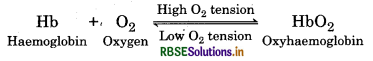
A haemoglobin molecule can be associated with upto four O2 molecules.
2. Transport of Carbon dioxide: In cellular respiration CO2 and water are released. Released CO2 diffuses into blood and combines with different blood components to produce various new products. These products carry CO2 to lung and release it into lung. The transport of CO2 in blood occurs in the following three main ways:
- In the form of carbonic acid (H2CO3).
- In the form of bicarbonates.
- In the form of carbamino compound.
Question 4.
Write short notes on inspiration and expiration.
Answer:
Inspiration: The entry of oxygen rich air into lungs is called inspiration. It is possible when the pressure of air in lungs is less than the atmospheric pressure. It includes following actions:
- First of all the radial muscles of diaphragm contract whereby diaphragm becomes flat.
- Now contraction occurs in external intercostal muscles, whereby ribs rise outwards and sternum rises upwards.
- As a result of both these actions the volume of thoracic cavity increases and lungs get expanded. In this way due to increase in the volume of lungs, the air pressure inside the alveoli becomes 1 to 3 mm Hg lesser than the atmospheric air pressure. Thereby air enters into the lungs. The path of air is as follows:


- Breathing, normally is an involuntary activity. It is also an active process in which energy is required.
- Expiration: The movement of carbon dioxide rich air from lungs to external atmosphere is called expiration. It is possible when the pressure of air in lungs is more than atmospheric pressure.
It includes following actions:
- There relaxation occurs in external intercostal muscles whereby ribs and sternum comes to their normal positions.
- Now relaxation occurs in radial muscles of diaphragm and it becomes dome shaped.
- As a result of both these actions, the volume of thoracic cavity reduces and the pressure increases on lungs. In this way, the air pressure inside the alveoli becomes 1 - 3 mm Hg more than the atmospheric air pressure.
- Hence air is expelled out through the respiratory tract.
- During normal breathing, the process of expiration is a passive process. In normal expiration no any muscle contraction occurs. After completion of the inspiration, the above mentioned respiratory muscles get relaxed thereby allowing the ribs and diaphragm to comes to their normal position. During exercise this turns as active.
The normal breathing rate of human is 12 - 16 per minute. But during exercise and flutter, the breathing rate increases. At this time, the contraction rate of muscles of diaphragm and intercostal muscles increases 4 - 5 times, thereby the volume of thoracic cavity also increases 15 - 20% in normal condition. By this respiratory capacity increases and cells, tissues and muscles can get oxygen.
Question 5.
Write short notes on the following:
(a) Abdominal breathing
(b) Forced breathing.
Answer:
(a) Abdominal breathing: It is calm breathing which drives mainly by movements of diaphragm. During expiration, flattening of diaphragm, a pressure is creates on abdominal organs. Due to this abdominal organs also create pressure on abdominal wall, thereby abdomen swells. It becomes normal in expiration. In this swallowing and flattening of thorax is much less.
(b) Forced Breathing: It is also called deep breathing. During this the intercostal muscles and abdominal muscles also contract which greatly reduces the volume of thoracic cavity thus exerting additional pressure on the lungs. The contraction of internal intercostal muscles decreases the volume of thoracic cavity by pulling the ribs and sternum dorsally and downward whereas the contraction of abdominal muscles causes abdominal organs to press the diaphragm upward which in turn bring about additional pressure on lungs as a result of which more than normal amount of air is thrown out.
During prolonged exercise, exhaustion or nervousness we take deep breath which is a voluntary activity. During this time, not only diaphragm gets fully contracted but external intercostal muscles also undergo complete contraction which increases the volume of thoracic cavity 20 - 25% more than during the usual breathing and so 20 - 25% more air is filled in the lungs.
Question 6.
Differentiate between inspiration and expiration.
Answer:
|
Inspiration |
Expiration |
|
1. In this atmospheric air enters into the lungs. |
1. In this lung air is expelled out. |
|
2. The air pressure in lungs becomes lesser during inspiration. |
2. The air pressure in lungs becomes greater during expiration. |
|
3. The radial muscles of diaphragm contract thereby diaphragm becomes flat. |
3. The radial muscles of diaphragm get relaxed thereby diaphragm becomes dome shaped. |
|
4. External intercostal muscles and cartilagenous part of internal intercostal muscles contract thereby thoracic cage pulled outwardly. |
4. Due to contraction of nternal intercostal muscles and relaxation of external intercostal muscles, thoracic cage pulled downwardly. |
|
5. The volume of pleural cavity is increased. |
5. The volume of pleural cavity is decreased. |
Question 7.
Write short notes on the following:
(i) Whooping cough
(ii) Common cold.
Answer:
(i) Whooping cough: This disaese in children is caused by Haemophilus pertussis. It is also a contaminating disease. In it patient feels cough and its effect becomes aggravated in night. It is called whooping cough.
(ii) Common Cold: This disease is caused by many viruses. Mucous membrane of nostrils get swallen and nose becomes closed, more secretion of mucous, asphyxiation, headache and. fever are many symptoms. This disease occur in people which have more age than 45 years or in children.
Long Answer Type Questions
Question 1.
What do you understand by aerobic anc anaerobic respiration? Give their examples.
Answer:
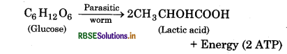
Respiration occuring in the absence of oxygen is called anaerobic respiration. During this process, the food substances are incompletely oxidised resulting in the formation of ethyl alcohol and CO2 from one molecule of glucose. The energy released during this respiration is quite less in comparison to aerobic respiration. The process of anaerobic respiration can be represented by the following reaction:
Anaerobic respiration also takes place in deep tissues of animals. It also can be seen in parasites of intestine such as tapeworm, round worm, hook worm and liver worm.
During anaerobic respiration, lactic acid is formed in muscles. Accumulating in muscles it causes muscle fatigue. Later, it gradually oxidise completely by liver cells and cardiac muscles. There is no mitochondria in red blood corpuscles therefore, anaerobic respiration in them also takes place.
Gaseous Exchange in Aerobic Respiration
All the aerobic organisms obtain oxygen for respiration from atmosphere by diffusion and release CO2. Air contains 21% and water contains 0.7% oxygen. Thus more oxygen is available for terrestrial organisms than that of aquatic organisms. This exchange of gases in organisms is of two types:
1. Direct gaseous exchange: In this direct exchange of O2 and CO2 takes place between cells of organisms and aquatic medium. There is no any transport medium for gaseous transport like blood. This type of exchange occurs in unicellular organisms such as animals of phylum protozoa and coelenterata.
2. Indirect gaseous exchange: In mostly multicellular organisms, there is no direct connection between somatic cells and external environment. They have specific respiratory organs. Therefore, the gaseous exchange occurs at two levels in these organisms:
(i) External Respiration: In this gaseous exchange takes place between external environment (water or air) and blood. This process is taken place at body surface, at respiratory surface and in respiratory organs. It includes breathing also. It is only a physical process.
(ii) Internal Respiration: In this gaseous exchange takes place between blood and tissue cells. This process is done at cellular level. It is also called tissue respiration. It includes gaseous exchange, oxidation of food and energy releasing also. Therefore, it is a physio - chemical process.
Question 2.
Give a brief description of respiratory organs in different organisms.
Answer:
Different types of respiratory organs are found in various organisms for gaseous exchange. The animals of phylum protozoa, porifera and coelenterata do not have specific respiratory organs. In these animals, the exchange of gases occurs through general surface by diffusion. The diffusion can takes place. through skin in frog. In earthworm, tapworm, round worm etc, the diffusion takes place through moist skin.
The animals of phylum arthropods have gills, trachea or book lungs for gaseous exchange. The gaseous exchange in mosty aquatic vertebrates is done by gills. Gills have vasculatory system and it has more blood vasculation thereby O2 dissolved in water is absorbed and CO2 is released in water. It is known as aquatic respiration. In terrestrial animals, the gaseous exchange is done by lungs. In which oxygen is obtained from atmosphere. Thus, it is known as aerobic respiration. Examples: Amphibians, reptiles, birds and mammals.
1. Respiration through general body surface: In the animals of phylum protozoa, porifera and coelenterata and many other animals which are living in water and wet environment, the respiration occurs by general body surface. These animals of phylum protozoa, porifera and coelenterata neither have specific respiratory organs nor circulatory system of gaseous transportation, in such condition, general wet body surface acts as gaseous exchange organ. But on the skin of earthworm and frog has moisture alongwith blood circulation which makes easy the gaseous exchange.
2. Respiration through gills: Gills are the main respiratory organs in some aquatic arthropods, molluscs and all the fishes. Gills has many micro - filaments, which are called gill filaments. They are surrounded by highly vascularised epithelium. When water pass through these filaments, then due to different concentration, the O2 moves into blood and CO2 comes out from blood to water and gaseous exchange takes place by simple diffusion. The direction of water flow and blood circulation in gills is opposite thereby a reverse flow system is formed which makes gaseous exchange easy.
3. Respiration through trachea: Due to lack of haemoglobin, the blood of insects does not function as oxygen carrier. Therefore, there is a network of trachea spreads in tissues and different parts of body in these animals for oxygen supply by 10 pairs of spiracles present laterally. Oxygen enters in these trachea. Trachea opens into atrium. Spiracles have bristles that filter the air. Each spiracle has valve to control opening and closing it.
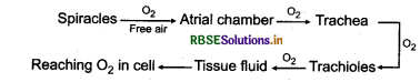
4. Lungs: The respiration takes place in amphibians, reptiles, birds and mammals through lungs. In this chapter, respiration process by lungs is described in detail.
Question 3.
Explain human respiratory system.
Answer:
Respiratory System of Human:
The main respiratory organ of human is lung and other organs serves as accessory organs. Respiratory system of human being consists of the following parts:
- Nose and nasal passage
- Pharynx
- Larynx
- Trachea
- Lungs
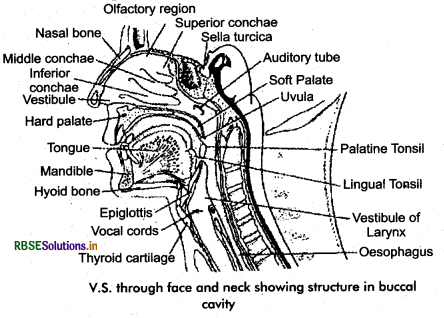
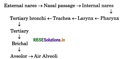
1. Nose and Nasal Passage
The respiratory system of human beings starts from nose. Nose is a hollow organ above the mouth, made of bone, cartilage, muscles and connective tissues. The cavity inside the nose is called nasal cavity which is divided by a nasal septum into right and left nasal passages. Both the nasal passage open outside in the form of two oval opening called external nares, or nostrils. Similarly, these nasal passages open posteriorly near glottis in the nasopharyngeal portion of pharynx. These openings are called internal nares. The roof of the nasal cavity consits of ethmoid, sphenoid and frontal bones whereas its floor is made of hard and soft palates which separate the nasal cavity from the mouth cavity. The lateral walls of the nasal cavity are made of maxilla, palatine, ethmoid and turbinate bones. The anterior portion of nasal septum is made of hyaline cartilage whereas the posterior portion is made of perpendicular plate of ethmoid bone and vomer.
Each nasal passage is divided into three parts:
(i) Vestibule: Vestibule is the small portion lying just behind the external nares. This portion of nose is lined by stratified squamous epithelium which is provided with stiff hairs which remove the dust particles coming with the inspired air.
(ii) Olfactory region: Olfactory region is the portion starting from the middle of the roof of the nasal cavity to 8 - 10 mm downward on the nasal septum and on the surface of superior conchae. This portion is lined by olfactory epithelium or Schneiderian membrane in which organ of sense of smell are situated.
(iii) Respiratory region: On the lateral walls of the nasal cavity, these three curved bony plates called superior, middle and inner conchae are present. The superior conchae are the elevations of ethmoid bone whereas the inner concha is a bone in itself and is also called turbinate bone. In such a way, these conchae divide each nasal passage into three : the superior, middle and lower air meatuses and thereby increase the surface area of the mucous membrane due to which the air passing from there becomes more hot and moist. The middle and lower conchae of the respiratory portion are lined by pseudostratified ciliated columnal epithelium which is provided with goblet cells here and there.
1. Function of Nose: Although we can breath with both nose and mouth but there are following benefits of breathing by nose:
- Being convoluted the nasal passage by turbinal bones their inner surface area is increased more. When hot air passes through these long paths becomes cold as body temperature.
- Nasal passages acts as filter and they filter air coming from outer side having dust particles and microorganisms. These enmesh in mucous and retained in nasal passages.
- Mucosa keeps nasal chambers moist thereby the air reaching into lungs becomes moist.
- Schnederian membrane has olfactory sense.
2. Phaiynx: The buccal cavity opens posteriorly into a funnel shaped cavity through internal nares, this cavity is called pharynx. It is 12 - 14 cm long muscular tube like structure. It may be divided into three parts:
(i) Nasopharynx: It is the portion of pharynx which is situated behind the nasal cavity and above the soft palate. A pair of internal nares and a pair pores of eustachin tube opens into it. A pair of protuberances made of lymphoid tissue are found hanging from the posterior part of the roof of nasopharynx. Internal nares are related to respiration and the eustachian pores related to tympanum.
(ii) Oropharynx: The part of pharynx situated just behind the buccal cavity is oropharynx. It extends posteriorly upto epiglottis from where respiratory and digestive passages get separated in the form of trachea and oesophagus respectively. On the anterio - lateral walls of the oropharynx also, protuberances of lymphoid tissues are found which are known as palatine tonsils.
(iii) Laryngopharynx: The posterior part of pharynx extending from the level of hyoid bone to the region behind the larynx is called laryngopharynx.
Function: The food and air reaches to food pipe and wind pipe respectively passing through oesophagus. During inspiration epiglottis move from glottis but at the time of deglutination soft palate rises upwardly and epiglottis again covers the glottis, thereby food can not reach into larynx. However the food particles reach into wind pipe, it would be cause rapid cough.
3. Wind pipe: Wind pipe enters thoracic cavity through neck. Wind pipe is situated ventral side to oesophagus. The wind pipe is divided into two portions. Upper chamber like portion called larynx situated just behind eipglottis. Lower long tubular portion called trachea.
The vocal cords are present in larynx. When air passing out through larynx them vibrations produce in vocal cords thereby sound produces.
4. Trachea: It is a thin walled elastic tube, starts from the lower end of larynx and extends upto the apex of lungs where it divides into two branches; the left and right bronchi which enter into each lung. The framework of trachea is made up of 16-20 C-shaped incomplete rings made of hyaline cartilage which surrounds the trachea from ventral and lateral sides and are mutually attached with the help of fibro-elastic connective tissues and involuntary muscle fibres. The dorsal side of trachea which is close to the oesophagus is devoid of cartilage but has a thick covering of longitudinal smooth muscles.
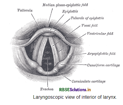
Larynx and Sound Production System
Larynx is a slender, irregular tube like structure, situated in front of 4th, 5th and 6th cervical vertebrae in the upper neck region, below the hyoid bone and the lower part of the tongue. Posteriorly it opens into trachea of the level of seventh cervical vertebra. The larynx of human is made of 9 cartilages. All these cartilages are movable and are mutually joined by ligaments. Cartilages are unpaired and paired. Unpaired cartilages: These are three:
(i) Thyroid cartilage: This is largest hyaline cartilage and forms ventro-lateral part of larynx. The thyroid cartilage of man is protruted frontly and looks triangular. This elevated portion of neck is called Adam’s apple. In women it is small in size. Anteriorly the thyroid cartilage is attached with hyoid bone, at the base of tongue with the help of ligament called thyrohyoid ligament.
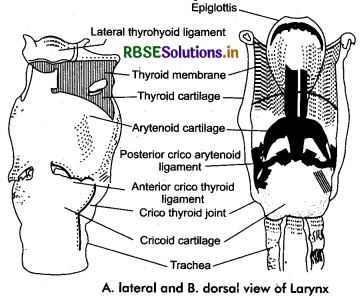
(ii) Epiglottis: It is a leaf like structure made of yellow elastic cartilage and is attached with hyoid bone and thyroid cartilage with the help of ligaments. It obliquely ascends upwards upto the region behind the tongue and above larynx. Its main function is to cover up the glottis during swallowing hence called epiglottis. It also covers up the oesophagus while inspiration.
(iii) Cricoid cartilage: It forms the base of larynx. It is a ring like cartilage situated below the thyroid cartilage. Cricoid cartilage is attached in the front portion with thyroid cartilage through cricothyroid ligament whereas it is attached again with the first cartilagenous ring of the trachea through cricotracheal ligament.
Paired Cartilages
- Arytenoid cartilage: Both of these cartilages are small and pyramid like made of hyaline cartilage which forms the dorsal wall of. larynx. Ligaments and muscles of vocal cords are attached with these cartilages.
- Corniculate cartilages: Both of these are small horn shaped cartilages which are attached with the upper ends of arytenoid cartilage.
- Cuneiform cartilages: Both of these are long and narrow, slender whedge shaped cartilage being covered by corniculate cartilage.
Vocal cords: The cavity of larynx or sound box is also known as laryngeal chamber. There are two pairs of vocal cords extend transversely between thyroid and arytenoid cartilages in laryngeal chamber. There are a pair of false vocal cords and a pair of true vocal cords in laryngeal chamber.
(i) False vocal cords: A pair of vocal cords which are slightly thick and less elastic, extend between thyroid and arytenoid cartilages in upper portion of laryngeal chamber. They do not take part in sound production. They function as to make moist the true vocal cords and support them.
(ii) True vocal cords: They are one pair, relatively thin, more elastic and white cords and situated beneath the false vocal cords in lower portion of laryngeal chamber. These both. extend between thyroid and arytenoid cartilages. The space between both true vocal cords is called rimaglottidis.
Question 4.
Describe sound producing organs of human.
Answer:
Sound Production: When the position of arytenoid cartilage is changed due to contraction of internal muscles of larynx then both the true vocal - cords bring closer so that the space between them is narrowed and rimaglottidis is formed. With the rapid passing of the air during expiration, vocal cords vibrate to produce sound. Due to relaxation of laryngeal muscles arytenoid.
muscles turn outwardly thereby widening the gap between vocal folds and the fissure and hence the sound production is ceased. Pitch of sound depends upon the length of folds and stress brought about in them which causes vibration in the folds. The change in voice in the form of different words depends upon the movement of the msucles of lips, tongue and soft palate. In boys the larynx is more developed than that of girls. As a result during puberty the voice in boys becomes heavy and in girls it becomes sharp.
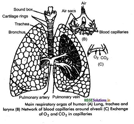
Lungs: In human two big conical lungs are main respiratory organs which are situated on both sides of the midline. The major portion of the thoracic cavity is occupied by the lungs. The space between both lungs is called mediastinum in which the heart, major blood vessels, trachea, oesophagus, thoracic duct and thymus gland are located. Each lung is pinkish coloured, dense and spongy structure and covered by a double layered serous membrane which is known as pleural (pleura membrane). The inner layer of pleura which is in contact with lungs is called visceral pleura and the outer layer which forms the inner coat of thoracic wall and also covers the upper surface of diaphragm is called parietal pleura. The space between both the layers of pleura is known as pleural cavity. This cavity is filled with serous fluid which keeps both the layers moist and smooth as a result of which these layers glide oyer each other. As a result of the inflammation of pleural cavity, the amount of serous fluid increases which results into a disease called pleurisy.
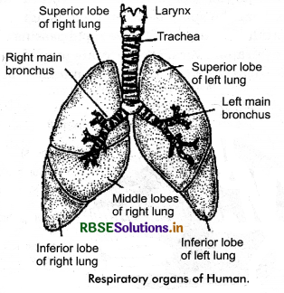
Each lung is divided into lobes by deep fissures. Right lung is divided into three lobes, superior, middle and inferior lobes by two oblique fissures. Left lung is divided into two lobes a larger superior lobe and smaller inferior lobe by an oblique fissure. The apical portion of each lung lies just above the clavicle bone whereas its basal portion is situated on the diaphragm. The protruding outer surfaces of lungs lie adjacent to the ribs and the muscles between them. The surface of lung facing mediastinum is concave on which a triangular region is present at the level of 5th to 7th thoracic vertebrae, which is known as hilus. It is the hilus through which bronchi, blood vessels, lymphatic vessels and nerves enter into lungs and come out from them.
Question 5.
Describe internal structure of human lung.
Answer:
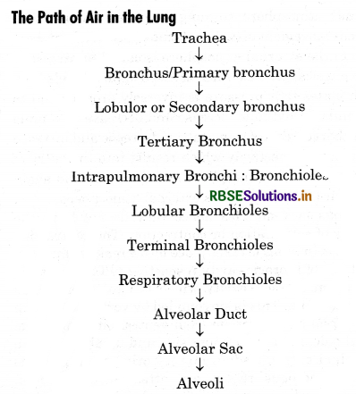
Trachea, reaching in thoracic region, divides into two main branches, known as primary bronchus. Each primary bronchus enters into each left and right lung through the hilus. The primary bronchus entering into left lung divides into two secondary bronchi which enter into the superior and inferior lobes. Similarly the primary bronchus which goes to right lung divides into three secondary bronchi, each of which enters into superior, middle and inferior lobes. These lobular bronchi of both sides redivide to form intrapulmonary bronchi.
There C shaped cartilagenous rings are found on trachea, bronchi, lobular bronchi and intrapulmonary bronchi whereby these do not collapse on empty. Next to them rings are absent on tubules. Each intra pulmonary bronchus divides to many ringless bronchioles. These bronchioles are divided into terminal bronchioles and each terminal bronchus divides into many respiratory bronchioles.
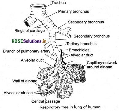
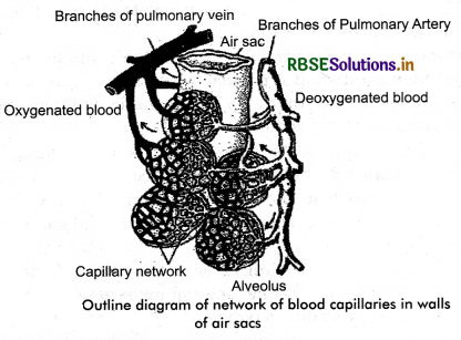
From each respiratory bronchiole 2 to 11 alveolar ducts originates each of which open into alveolar sacs, made of two or three sacs like alveoli. The space present between the alveolar duct and alveolar sac is known as atrium. These alveolies are smallest structural and functional units of lung. These are made of squamous epithelial layer and there dense network of blood capillaries found around them. Pulmonary artery carry deoxygenated blood into alveolies and pulmonary vein carry out oxygenated blood from them.
It is clear from the above description that air tubes undergo branching. So many times between their entry into lungs to their final arrival in the alveoli and thereby form a tree like structure known as bronchial tree. Bronchial tree has been divided into two zones, conducting zone and respiratory zone. The portion from trachea to the terminal bronchioles is called conducting zone which is used for conduction of air. The portion of bronchial tree beyond the terminal bronchioles to the alveoli is termed as respiratory zone where exchange of gases between air and the blood is accomplished. Approximately 60 crores of alveoli are present in both the lungs, which have an area of 100 square meters. Each alveoli is a thin walled bag like structure which is made of two fine layers.
The inner layer made of squamous epithelium and the outer layer made of connective tissues. A dense network of blood capillaries present in the connective tissue layer which get bulged in the alveoli so that their maximum surface area is exposed to the air of alveoli. In addition to the blood capillaries reticular and elastic fibres, lymphatic capillaries and macrophages are also present in the connective tissues. A thin layer of mucous is also present on inner and outer layer of alveoli. The surface of alveolus is called respiratory surface. Which is 0.2 mm in diameter.
Question 6.
Give brief description of breathing.
Answer:
Pulmonary Ventilation:
The exchange of air between external atmosphere and lungs alveoli is called pulmonary ventilation or breathing. The movement of oxygen (O2) rich air from external atmosphere to lung is called inspiration whereas movement of carbon dioxide (CO2) rich air from lungs to the external atmosphere is called expiration. It is a physical process which is based upon Boyle’s law, which states that pressure is inversely proportional to the volume. Rhythmic increase and decrease in volume of the thoracic cavity brings about decrease and increase in pressure respectively which results into the entry of air from external atmosphere into the lungs and again exit of air from lungs to the external atmosphere.
The lungs lack muscles hence they do not have the capacity of self dilation or contraction. The contraction or dilation in lungs is takes place by decrease or increase in volume of thoracic cavity respectively. The lungs are situated in thoracic cavity which is like a box, called thoracic cage. This is surrounded by vertebral column from dorsal side, sternum from ventral side, ribs from lateral side, neck from upper side and diaphragm from abdominal cavity.
Radial muscles originating from its central tendinous region are attached to sternum, thoracic vertebrae, posterior ribs and abdominal wall. In normal condition, diaphragm remains dome-shaped whose elevated portion faces the thorax region. There is 12 pairs of ribs present in the lateral region of thoracic cavity. On the dorsal side head of each rib is attached with the main body of the vertebra and the tubercle is attached with the lateral process of the vertebra whereas on the ventral side it is attached to the sternum with the help of cartilage. Ribs of both the sides are mutually joined by oblique striated intercostal muscles. These muscles are of two types: External Intercostal Muscles = EICM and Internal Intercostal Muscles = IICM.
A pair intercostal muscles present between two sequential ribs. In this way, 11 pairs of intercostal muscles are present in between 12 pairs of ribs.
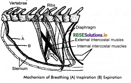
The movement of ribs occurs due to contraction and relaxation of these muscles. In ventilation diaphragm and radial muscles play 75% role and intercostal muscles of ribs play 25% role. The thoracic cage or thoracic box is an air tight chamber. The increase or decrease in volume of thoracic cavity and lungs occur by increase or decrease in volume of thoracic box. If diaphragm or respiratory box can be punctured then thoracic cavity unable to cause pressure on lungs and breathing will stopped and within a few time the person will die.
Mechanism of Breathing: It is completed in two steps: Inspiration and expiration.
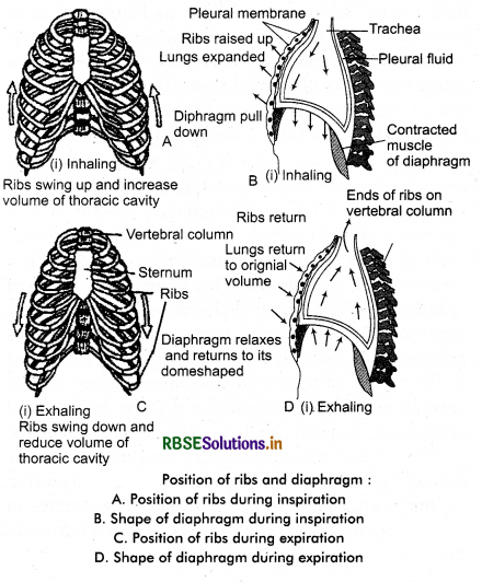
Inspiration: The entry of oxygen rich air into lungs is called inspiration. It is possible when the pressure of air in lungs is less than the atmospheric pressure. It includes following actions:
- First of all the radial muscles of diaphragm contract whereby diaphragm becomes flat.
- Now contraction occurs in external intercostal muscles, whereby ribs rise outwards and sternum rises upwards.
- As a result of both these actions the volume of thoracic cavity increases and lungs get expanded. In this way due to increase in the volume of lungs, the air pressure inside the alveoli becomes 1 to 3 mm Hg lesser than the atmospheric air pressure. Thereby air enters into the lungs. The path of air is as follows:
(iv) Breathing, normally is an involuntary activity. It is also an active process in which energy is required.
Expiration: The movement of carbon dioxide rich air from lungs to external atmosphere is called expiration. It is possible when the pressure of air in lungs is more than atmospheric pressure. It.includes following actions:
- There relaxation occurs in external intercostal muscles whereby ribs and sternum comes to their normal positions.
- Now relaxation occurs in radial muscles of diaphragm and it becomes dome shaped.
- As a result of both these actions, the volume of thoracic cavity reduces and the pressure increases on lungs. In this way, the air pressure inside the alveoli becomes 1 - 3 mm Hg more than the atmospheric air pressure.
- Hence air is expelled out through the respiratory tract.
- During normal breathing, the process of expiration is a passive process. In normal expiration no any muscle contraction occurs. After completion of the inspiration, the above mentioned respiratory muscles get relaxed thereby allowing the ribs and diaphragm to comes to their normal position. During exercise this turns as active.
The normal breathing rate of human is 12 - 16 per minute. But during exercise and flutter, the breathing rate increases. At this time, the contraction rate of muscles of diaphragm and intercostal muscles increases 4 - 5 times, thereby the volume of thoracic cavity also increases 15 - 20% in normal condition. By this respiratory capacity increases and cells, tissues and muscles can get oxygen.
(a) Abdominal breathing: It is calm breathing which drives mainly by movements of diaphragm. During expiration, flattening of diaphragm, a pressure is creates on abdominal organs. Due to this abdominal organs also create pressure on abdominal wall, thereby abdomen swells. It becomes normal in expiration. In this swallowing and flattening of thorax is much less.
(b) Forced Breathing: It is also called deep breathing. During this the intercostal muscles and abdominal muscles also contract which greatly reduces the volume of thoracic cavity thus exerting additional pressure on the lungs. The contraction of internal intercostal muscles decreases the volume of thoracic cavity by pulling the ribs and sternum dorsally and downward whereas the contraction of abdominal muscles causes abdominal organs to press the diaphragm upward which in turn bring about additional pressure on lungs as a result of which more than normal amount of air is thrown out.
During prolonged exercise, exhaustion or nervousness we take deep breath which is a voluntary activity. During this time, not only diaphragm gets fully contracted but external intercostal muscles also undergo complete contraction which increases the volume of thoracic cavity 20 - 25% more than during the usual breathing and so 20 - 25% more air is filled in the lungs.
Question 7.
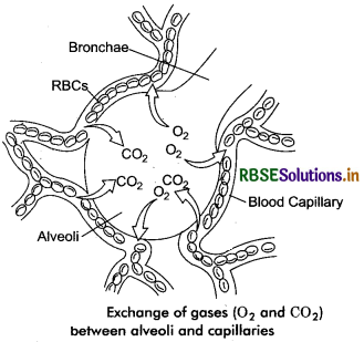
Explain gaseous exchange in human.
Answer:
There are about 60 crores alveoli found in human lungs which provide a total surface area of about 100 square metre which is about 50 times the surface area of the body. The walls of alveoli are very thin and made of squamous epithelium. These are permeable for both O2 and CO2. There is a dense network of blood capillaries around the walls of alveoli. The epithelium of alveoli and endothelium of blood capillaries comes closer to form a respiratory membrane. It is about 0.2 pm thick. The exchange of respiratory gases between blood of capillaries and air of alveoli occurs across this membrane by simple diffusion.
The trachea, bronchus, bronchioles and alveolar ducts lack blood capillaries, therefore no gaseous exchange occurs in other part except alveoli. There is a limit of diffusion of gases between alveoli and blood across the respiratory membrane which is known as diffusing capacity and is defined as the volume of gases being diffused across the membrane per minute at the difference of 1 mm Hg of pressure. At a specific pressure CO2 diffuses 20 times more rapidly in comparison to O2 whereas O2 diffuses 2 times more rapidly as compared to N2.
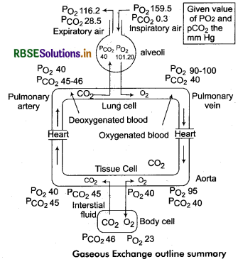
During a normal breathing about 500 mL of air is inspired and expired, out of which about 140 mL fills the dead space and rest 360 mL is available for gaseous exchange in the alveoli. The inspired air when passes through the nasal passage, it becomes saturated with water due to which concentration of CO2 present in air is not very much affected but the oxygen concentration reduces from 20.96% to 19.67%.
The partial pressure of O2 (PO2) m alveoli is 100 - 104 mm Hg while the partial pressure of CO2 (PCO2 ) in alveoli is 40 mm Hg. In deoxygenated blood coming in blood capillaries of lungs, the partial pressure of O2 is 40 mm Hg and the partial pressure of CO2 is 45 - 46 mm Hg. The amount of O2 is more in alveoli coming from outer air. This oxygen diffuses into pulmonary cells dissolving in mucous present on moist walls of alveoli. CO2 present in blood capillaries coming from tissues diffuses into alveoli, In this way blood capillaries have oxygenated blood. Expelled air from lung has 15.7% O2 and 3.6% CO2.
|
Atmosphere |
|
Alveolar air |
|
Blood |
|
PO2 =159.5 mm Hg |
→ |
PO2 =1012 mm Hg |
→ |
PO2 =40mm Hg |
|
PCO2 =0.3 mm Hg |
← |
PCO2 =40 mm Hg |
← |
PCO2=46 mm Hg |
Question 8.
How O2 and CO2 are transported by blood in human?
Answer:
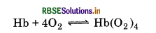
Oxygenated blood is carried from lungs to the left auricle by four pulmonary veins. From there it goes to left ventricle. There is it pumped out from the heart and all the body parts are supplied with the pure blood. Where the concentration of O2 is lesser and the concentration of CO2 is more, the oxyhaemoglobin dissociates into haemoglobin and oxygen.
Hb(O2)4 → Hb + 4O2
Released oxygen diffuses into tissue fluid and lymph through walls of blood capillaries and from there it reaches into tissue cells of organs. Inside the cells food substances are oxidised with the help of this oxygen. As a result CO2 and water are released. This CO2 is carried into lungs.
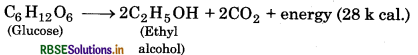
C6H12O6 + 6O2 → 6CO2 + 6H2O + 673 kcal
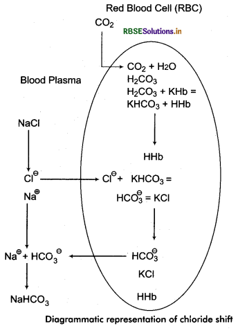
Transport of CO2 by Blood:
The CO2 is produced during oxidation of food substances stored in tissues. It moves into blood capillaries by diffusion. The transport of CO2 in blood occurs in the following ways:
1. In the form of carbonic acid: CO2 is highly soluble in water. About 10% of CO2 combines with water of plasma to form carbonic acid (H2CO3).
CO2 + H2O → H2CO3
About 10% part of total CO2 remains as H2CO3 in blood and rest part breaks down rapidly into hydrogen and bicarbonate ions.
H2CO3 → HCO3- + H-
2. In the form of Bicarbonate: About 70% of the CO2 is transported through the plasma in the form of bicarbonates of sodium and potassium.
CO2 +H2O ⇌ H2CO3 ⇌ HCO3- + H+
HCO3- + Na+ → NaHCO3 (Sodium bicarbonate)
HCO3- + K+ → KHCO3 (Potassium bicarbonate)
3. In the form of Carboxy haemoglobin: Some CO2 combines with haemoglobin of RBCs to form an unstable compound carboxyhaemoglobin.
Hb + 4CO2 → Hb(CO2)4
4. In the form of carbamino compound: About 7% CO2 combines with protein of plasma to form carbamino compound.
Plasma protein + CO2 → Carbamino compound (unstable)
The blood with carbonic acid, sodium and potassium bicarbonates, carboxyhaemoglobin and carbamino compound is impure blood. This blood goes from capillaries to heart through veins and from here to lungs through pulmonary arteries. From lungs CO2 diffuses to alveoli.
5. Release of CO2 from unstable substances: The amount of carboxyhaemoglobin is more in blood capillaries near the lungs. This is more acidic. Due to having acidic nature of carboxyhaemoglobin, all the unstable compounds break down from it and release CO2.
2NHCO3 → Na2CO3 + H2O + CO2
2KHCO3 → K2CO3 + H2O + CO2
H2CO3 → H2O + CO2
Hb(CO2 )4 → Hb + 4CO2
Such released CO2 reaches to lung alveoli by diffusion through walls of thin capillaries. From where it is expelled out by expiration.
Chloride Shift
Exchange of bicarbonate and chloride ions between red blood cells and plasma is called chloride shift or Hamburger’s phenomenon. The formation of carbonic acid in plasma occurs at extermely slow rate but it occurs about 5000 times faster inside the RBCs due to presence of carbonic anhydrase enzyme. Most of the CO2 is diffused into RBCs from the plasma. In such corpuscles it is changed into carbonic acid in the presence of carbonic anhydrase. Carbonic acid in such corpuscles is dissociated into bicarbonate ions (HCO3-) and hydrogen ions (H+ ). Most of the H+ ions are neutralised by the buffer action of haemoglobin whereas HCO3- ions comes outside from the RBCs in the plasma by diffusion. In plasma HCO3- associate with Na+ ions to form sodium bicarbonate (NaHCO3). To maintain the electrostatic neutrality of the plasma the same amount of chloride ions (Cl-) moves inside the RBCs to compensate the bicarbonate ion. It is called chloride shift.
Question 9.
Explain respiratory disorders of human.
Answer:
Following are main respiratory diseases:
1. Asthma: It is an allergic disease caused by pollen grains, dust, smoke, smoking etc. It leads difficulty in breathing. The main symptom of this disease is continuous coughing. In . this disease, bronchi get narrowed due to spasm in smooth muscles present in the walls of bronchi and there also occurs peroxysomes of breathlessness as a result of excessive secretion of mucous due to swelling in mucosal cells. There is also stiffness in chest followed by dyspnea (pain during breathing) and wheezing. The only measure to central this disease is to keep away from above mentioned allergens. In case of emergency bronchodilators, oxygen treatment and corticosteroids are suggested.
2. Bronchitis: In this disease, along with inflammation of bronchi and bronchioles, there is sharp increase in the number of goblet cells and hypertrophy of mucous glands and finally decrease in the number of ciliated cells. These features results into excessive secretion of mucous but inhibition of its transport in the respiratory passage. This causes blockage of air inside the air paths. For escape from this disease keep away smoking and polluted environment.
3. Emphysema: It is a long term progressive disease of the lungs that primarily causes shortness of breath due to overinflation of the alveoli. In people with emphysema, the lung tissue involved in exchange of gases is impaired or destroyed. These features results into the loss of elasticity of these portions as a result of which alveoli remain filled with air even after expiration. Late complications result into respiratory failure and right heart failure.
4. Silicosis and Asbestosis: Silicosis disease is caused by the entry of silica and silicon dioxide ladden dust particles into the lungs. Asbestosis is caused by entry of asbestos fibres dust with breathing. The possibility of these diseases is in such persons whose working in silica and asbestos, factories, found more.
5. Pneumonia: It is a lung infection by bacterium streptococcus pneumoniae. The alveoli filled with dead cells and fluid being infection in lungs. These have swelling thereby a difficulty in breathing occurs.
6. Dyspnea: In this disease the person feels an anxiety, breathing rate increases. It is usually caused during heavy exercise, running or due to fearness.

- RBSE Solutions for Class 11 Biology Chapter 10 Cell Cycle and Cell Division
- RBSE Solutions for Class 11 Biology Chapter 9 Biomolecules
- RBSE Solutions for Class 11 Biology Chapter 8 Cell: The Unit of Life
- RBSE Solutions for Class 11 Biology Chapter 7 Structural Organisation in Animals
- RBSE Solutions for Class 11 Biology Chapter 6 Anatomy of Flowering Plants
- RBSE Solutions for Class 11 Biology Chapter 5 Morphology of Flowering Plants
- RBSE Solutions for Class 11 Biology Chapter 4 Animal Kingdom
- RBSE Solutions for Class 11 Biology Chapter 3 Plant Kingdom
- RBSE Solutions for Class 11 Biology Chapter 2 Biological Classification
- RBSE Solutions for Class 11 Biology Chapter 1 The Living World
- RBSE Solutions for Class 11 Biology Chapter 5 पुष्पी पादपों की आकारिकी